project-details

What is MRI

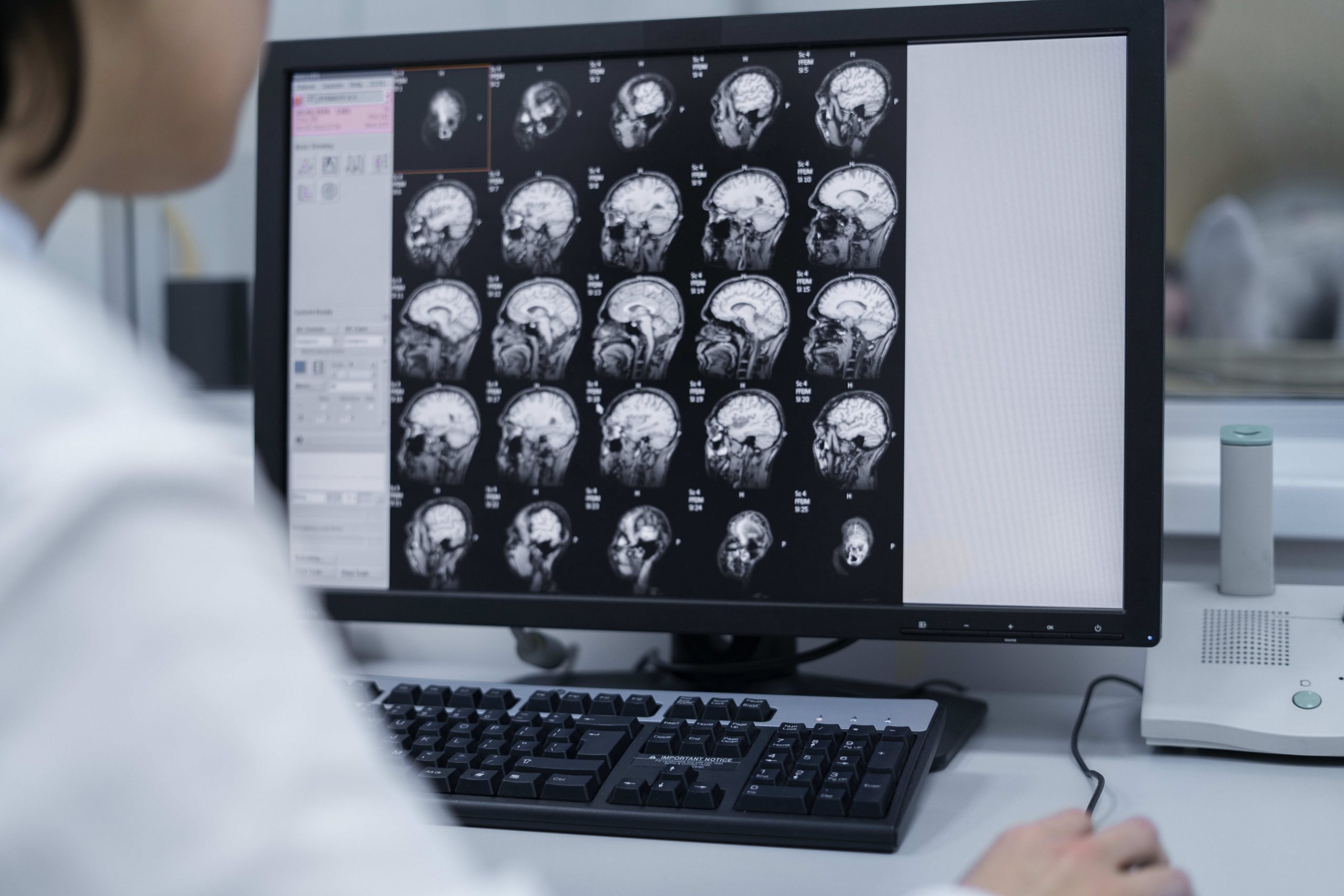
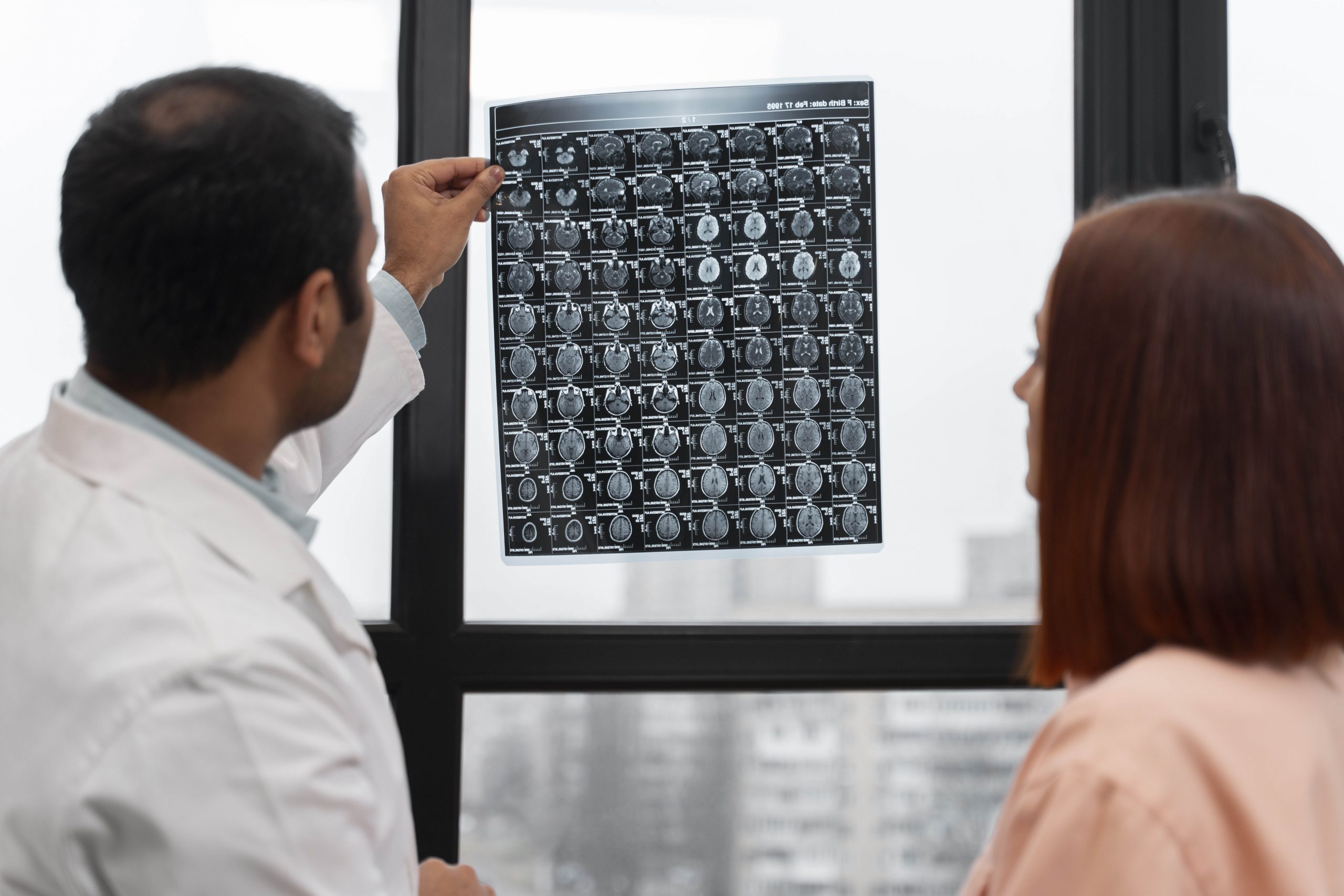
Magnetic resonance imaging (MRI) is a medical imaging technique used in radiology to form pictures of the anatomy and the physiological processes of the body. MRI scanners use strong magnetic fields, magnetic field gradients, and radio waves to generate images of the organs in the body. MRI does not involve X-rays or the use of ionizing radiation. This factor distinguishes it from CT and PET scans. It is the Gold Standard for imaging the brain and the spinal cord. Since its development in the 1970s and 1980s, MRI has proven to be a versatile imaging technique. MRI is most prominently used in diagnostic medicine and biomedical research.

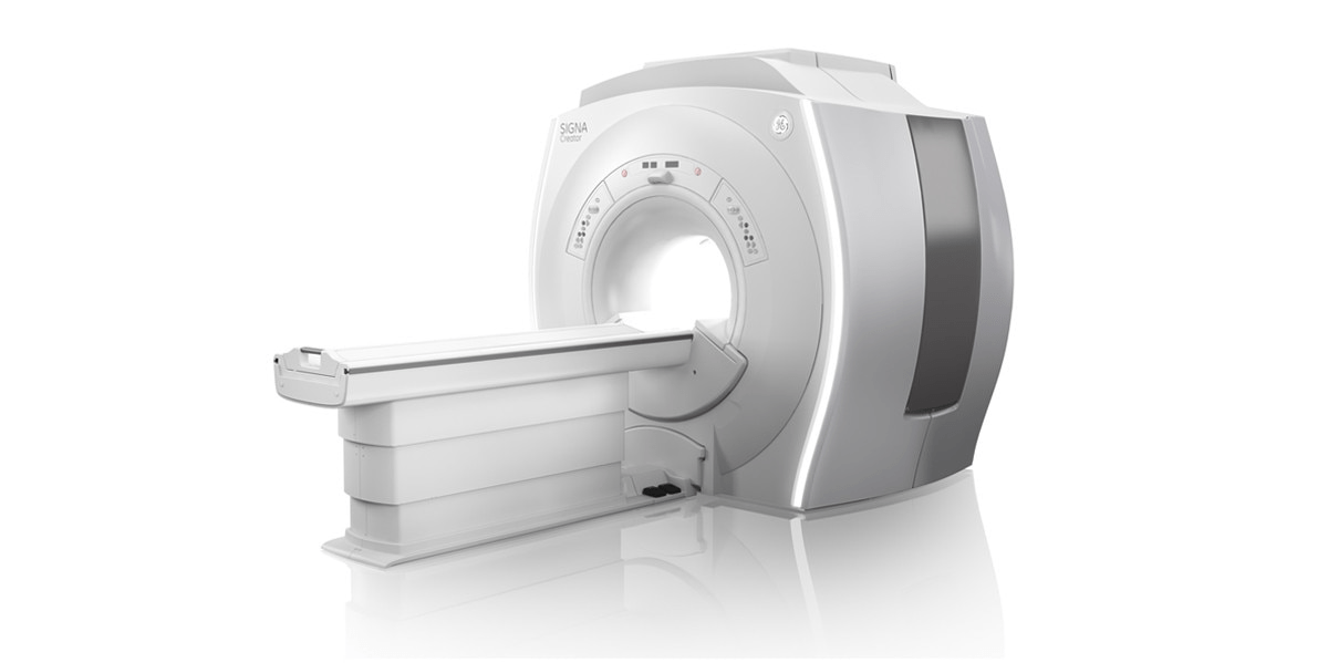
MRI Ge
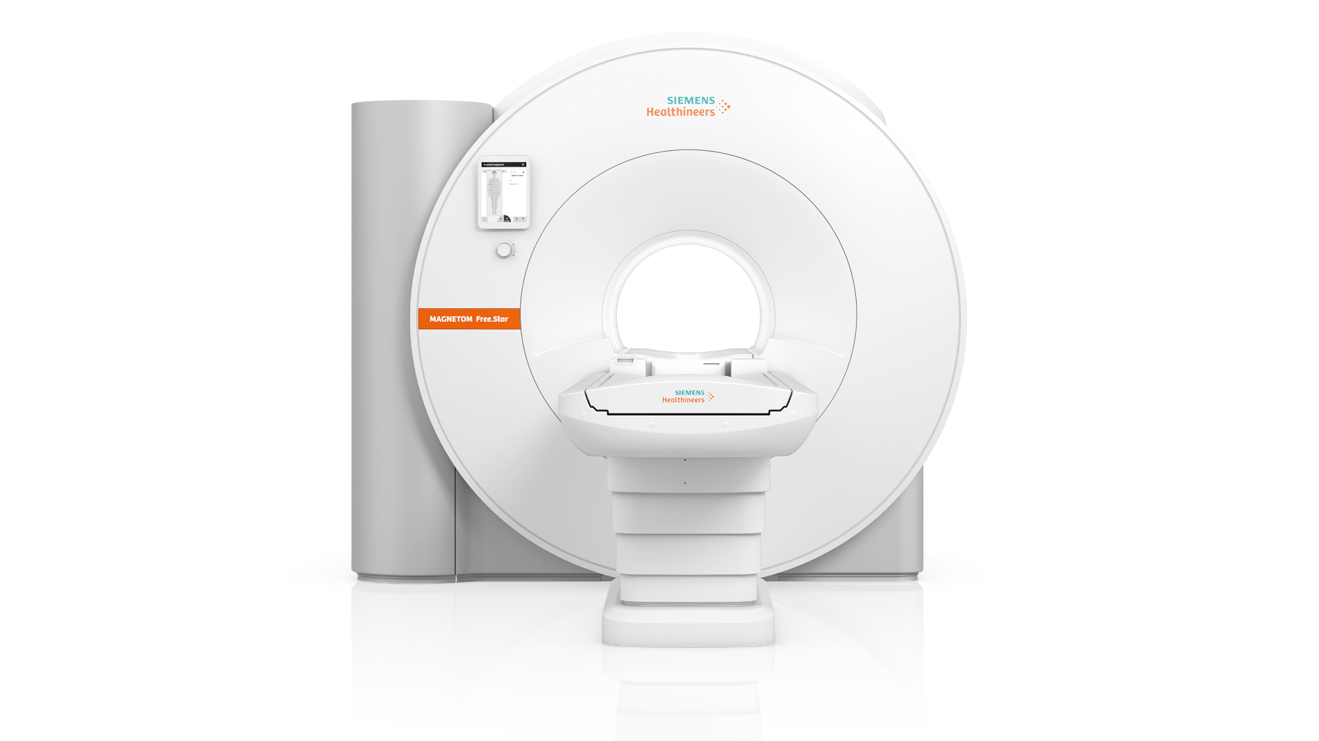
MRI Machine

Need for MRI Scan :

Types Of MRI Scans

Full Body

What can a full-body MRI scan tell you?

A full-body scan could show you’re in good health, giving you reassurance and helping you to plan a healthy future lifestyle. If there is a potential problem, the scan could identify a disease or condition at an early stage, contributing to effective treatment and a positive outcome.

icon

Head and brain

Your scan can identify many pathologies if they’re present, including brain tumours, evidence of a stroke or infarction, bleeds, white matter changes, and any presence of aneurysms.

icon

Abdomen and pelvis

Your full-body scan will look at major organs including the liver, kidneys, pancreas, and spleen, identifying any abnormalities such as tumours, lesions, and masses.

icon

Spine

The spine is scanned in three parts, including the lumbar, thoracic, and cervical regions, to identify any abnormalities or conditions affecting these areas. The scan accesses the intravertebral and the spinal cord, recognizing degenerative changes, disc herniation and prolapse, facet joint degeneration, and any spine cord lesions, or masses such as meningiomas.

icon

Heart (Platinum package)

Your cardiac MRI scan will assess the function and anatomy of your heart, looking for wall motion abnormalities, valve abnormalities and evidence of hypertrophy and myocardial infarction.

icon

Head and brain

Your scan can identify many pathologies if they’re present, including brain tumours, evidence of a stroke or infarction, bleeds, white matter changes, and any presence of aneurysms.

icon

Abdomen and pelvis

Your full-body scan will look at major organs including the liver, kidneys, pancreas, and spleen, identifying any abnormalities such as tumours, lesions, and masses.

icon

Spine

The spine is scanned in three parts, including the lumbar, thoracic, and cervical regions, to identify any abnormalities or conditions affecting these areas. The scan accesses the intravertebral and the spinal cord, recognizing degenerative changes, disc herniation and prolapse, facet joint degeneration, and any spine cord lesions, or masses such as meningiomas.

icon

Heart (Platinum package)

Your cardiac MRI scan will assess the function and anatomy of your heart, looking for wall motion abnormalities, valve abnormalities and evidence of hypertrophy and myocardial infarction.

Spine

What is a spine MRI scan?

MRI stands for Magnetic Resonance Imaging, which is a way of taking pictures inside the body using magnetic fields and radio waves. It’s a safe, painless and non-invasive procedure capturing high-quality images. Unlike X-rays, MRI doesn’t use potentially harmful radiation.

A spine MRI scan can focus on one of three defined areas of the spine, depending on your symptoms and where you’re experiencing pain.

What kinds of private spine MRI scans are available?

We provide a number of different types of spine MRI scans:

Cervical Spine Scan

This will include images of your C1 to C7 vertebrae - your neck bones. They go from the base of your skull to the top of your ribcage and are the most flexible parts of your spine.

Thoracic Spine Scan

This covers the 12 vertebrae in the middle of your spine, labelled T1 to T12. These vertebrae start at the base of your neck and go down to the bottom of the ribs.

Lumbar and Sacral Spine Scan

The lumbar spine comprises the final five vertebrae between your ribs and your pelvis. These vertebrae are designated as:
L1, L2, L3, L4, L5

It is the second most flexible section while also carrying the most weight, making it the most susceptible to injury. The sacrum (the bony ‘shield’ at the base of your spine) supports and connects the spine to the pelvis. It attaches to the pelvic girdle by the sacroiliac joints, forming part of the protection of the internal pelvic organs. It is a fusion of 5 vertebrae through which the lower sacral nerves pass into the pelvis.

What symptoms would prompt a spine MRI scan?

If you’re experiencing any of the following symptoms, a spine MRI scan could diagnose the problem and help you plan treatment.

What can a spine MRI scan detect? 

The results of your private MRI spine scan could help diagnose a range of injuries or conditions, including:

A spine MRI scan is a diagnostic tool, so it won’t treat these conditions. However, the results will allow your physician to make informed decisions and recommend appropriate treatment.

How do I prepare for a spine MRI scan?

Our patient care team will ensure you’re ready for your scan, but you can help by telling them about your medical history. Unless you’re asked to do otherwise, you can continue to eat, drink and take medication as normal before your scan.

Some implants and conditions may affect how your MRI scan is performed, or whether it’s safe to do it at all. These include

If any of these applies to you, please raise the issue with the patient care team before booking an appointment.

What happens during a spine MRI scan, and how long does it take?

Your radiologist will ask you to take off any jewellery and empty your pockets, or you may be asked to change into a gown. You’ll then lie on the MRI table during the scan. A single part spine MRI (such as Lumbar and Sacral scan) takes around 20 minutes, and we would ask you to arrive around 15 minutes before your scheduled appointment. 

Head

 What is a head MRI scan?

MRI stands for Magnetic Resonance Imaging, which is a way of taking pictures inside the body, including the head, using magnetic fields and radio waves. It’s a safe, painless and non-invasive procedure, capturing high-quality images. Unlike X-rays, MRI doesn’t use potentially harmful radiation.

MRI scanning technology is used extensively to examine the head, diagnosing and assessing a wide range of conditions a ecting the brain, eyes and inner ears.
Here we provide di erent types of MRI scans focused on di erent areas including the head & brain, inner ears and orbits (eyes).

What symptoms would prompt a head MRI scan?

A head MRI can help identify and assess a wide range of conditions associated with the brain, the eyes and eyesight, and the inner ears. Typical symptoms which might warrant a head MRI scan include:

Brain & Head MRI scan

A scan of the brain allows your radiologist to see if there are any issues in its structures, including:

Orbits (the eye area) MRI scan

Your orbits consist of several complicated structures associated with the eyes and sight. A scan will show:

Inner ears (IAMS) MRI scan

An inner ear scan helps diagnose problems within the internal auditory meatus (IAM), or inner ear canal. The images will show the bones, nerves, and other tissues in that area to diagnose a range of conditions, including hearing loss and tinnitus.

What can a head MRI scan detect?

Depending on the area being scanned, an MRI scan of the head can help diagnose and assess a wide range of diseases, conditions and injuries.

Brain & Head MRI scan

Your radiologist may diagnose you with

  • Bleeding
  • Inflammation or swelling
  • Fluid build-up
  • Brain development problems
  • Tumours
  • Infections
  • Pituitary disorders

Orbits scan

This could reveal the presence of

  • Haemorrhage
  • Inflammation or swelling
  • Trauma
  • Optic neuropathy

Inner Ears scan

Among the conditions this could diagnose are:

  • Acoustic neuromas
  • Sensorineural hearing loss
  • Meniere’s disease, and other conditions that cause balance problems
  • Tinnitus

How do I prepare for a head MRI scan?

Our patient care team will ensure you’re ready for your scan, but you can help by telling them about your medical history. Unless you’re asked to do otherwise, you can continue to eat, drink and take medication as normal before your scan.

Some implants and conditions may a ect how your MRI scan is performed, or whether it’s safe to do it at all. These include

If any of these applies to you, please raise the issue with the patient care team before booking an appointment.

What happens during a head MRI scan, and how long does it take? Your radiologist will ask you to take o any jewellery and empty your pockets, or you may be asked to change into a gown. You’ll then lie on the MRI table during the scan.

Knee

 What is a knee MRI scan?

MRI stands for Magnetic Resonance Imaging, which is a way of taking pictures inside the body using magnetic fields and radio waves. It’s a safe, painless and non-invasive procedure which captures high-quality images inside the body.
Unlike X-rays, MRI doesn’t use potentially harmful radiation.

During a knee joint MRI scan, the radiologist will use the scanner to take crosssection images of your knee showing the structures within it, including:

Why would I choose to have a private knee MRI scan?

You should consider getting an MRI scan of your knee if you’ve been experiencing problems that have lasted longer than a few weeks. Symptoms include

What can a knee MRI scan detect?

Your knee pain may be the result of an injury – from sport or a fall, for example – or could indicate another condition or problem. Some of the possible issues the scan can detect are

How do I prepare for a Knee MRI scan?

Our team will ensure you’re ready for your scan, but you can help them by telling them about your medical history. Unless you’re asked to do otherwise, you can continue to eat, drink and take medication as normal before your scan.

Some implants and conditions may affect how your MRI scan is performed, or whether it’s safe to do it at all. These include

If any of these applies to you, please raise the issue with the patient care team before booking an appointment.


What happens during a knee MRI scan, and how long does it take?

Your radiologist will ask you to take off any jewellery and empty your pockets, or you may be asked to change into a gown. You’ll then lie on the MRI table during the scan. A knee MRI scan takes around 20 minutes, and we would ask you to arrive around 15 minutes before your scheduled appointment.

Hip

What is a hip MRI scan?

MRI stands for Magnetic Resonance Imaging, which is a way of taking pictures inside the body using magnetic fields and radio waves. It’s a safe, painless and non-invasive procedure, which captures high-quality images. Unlike X-rays, MRI doesn’t use potentially harmful radiation.

During a hip MRI scan, the radiologist will use the scanner to produce detailed images of your hip’s inner structure, showing any damage or abnormalities in your

Why would I choose to have a private hip MRI scan?

If you’re experiencing any of these symptoms, you may want to arrange a private hip MRI scan.

What can a hip MRI scan detect?

Your hip MRI scan will detect damage to tissues and bones. Many different causes of hip pain can be identified during your hip MRI scan. They include:

How do I prepare for a hip MRI scan?

Our Patient Care Team will ensure you’re ready for your scan, but you can help by telling them about your medical history. Unless you’re asked to do otherwise, you can continue to eat, drink and take medication as normal before your scan.

Some implants and conditions may affect how your MRI scan is performed, or whether it’s safe to do it at all. These include

If any of these applies to you, please raise the issue with the patient care team before booking an appointment.


What happens during a hip MRI scan, and how long does it take?

Your radiologist will ask you to take o any jewellery and empty your pockets, or you may be asked to change into a gown. You’ll then lie on the MRI table during the scan. A hip MRI scan takes around 20 minutes, and we would ask you to arrive around 15 minutes before your scheduled appointment.

Hand & Wrist

 What is a hand and wrist MRI scan?

MRI stands for Magnetic Resonance Imaging, which is a way of taking pictures inside the body using magnetic fields and radio waves. It’s a safe, painless and non-invasive procedure, which captures high-quality images. Unlike X-rays, MRI doesn’t use potentially harmful radiation.

The hand and wrist area is complex, with many different bones and other structures, as well as different functions. Our hand and wrist MRI scanning service will focus on a particular area, depending on your symptoms.

Scans include:

Why would I choose to have a private hand or wrist MRI scan?

You should consider getting an MRI scan of your hand or wrist if you’ve been experiencing problems that have lasted longer than a few weeks.

Symptoms include:

What can a hand or wrist MRI scan detect?

This depends on the part of the hand and wrist area being scanned, which will be determined by your symptoms. The scans can detect 

Wrist scan 

  • Injuries (from impacts and repetitive stress)
  • Arthritis
  • Ganglion cysts
  • Kienbock’s disease
  • The severity of carpal tunnel syndrome

Palm scan 

  • Fractures
  • Inflammation
  • Ganglion cysts
  • Ligament tears
  • Nerve damage

Thumb or finger scan 

  • Fractures
  • Osteoarthritis
  • Tendinitis

How do I prepare for a hand and wrist MRI scan?

Our patient care team will ensure you’re ready for your scan, but you can help by telling them about your medical history. Unless you’re asked to do otherwise, you can continue to eat, drink and take medication as normal before your scan.  

Some implants and conditions may affect how your MRI scan is performed, or whether it’s safe to do it at all. These include 

If any of these applies to you, please raise the issue with the patient care team before booking an appointment. 

Abdomen

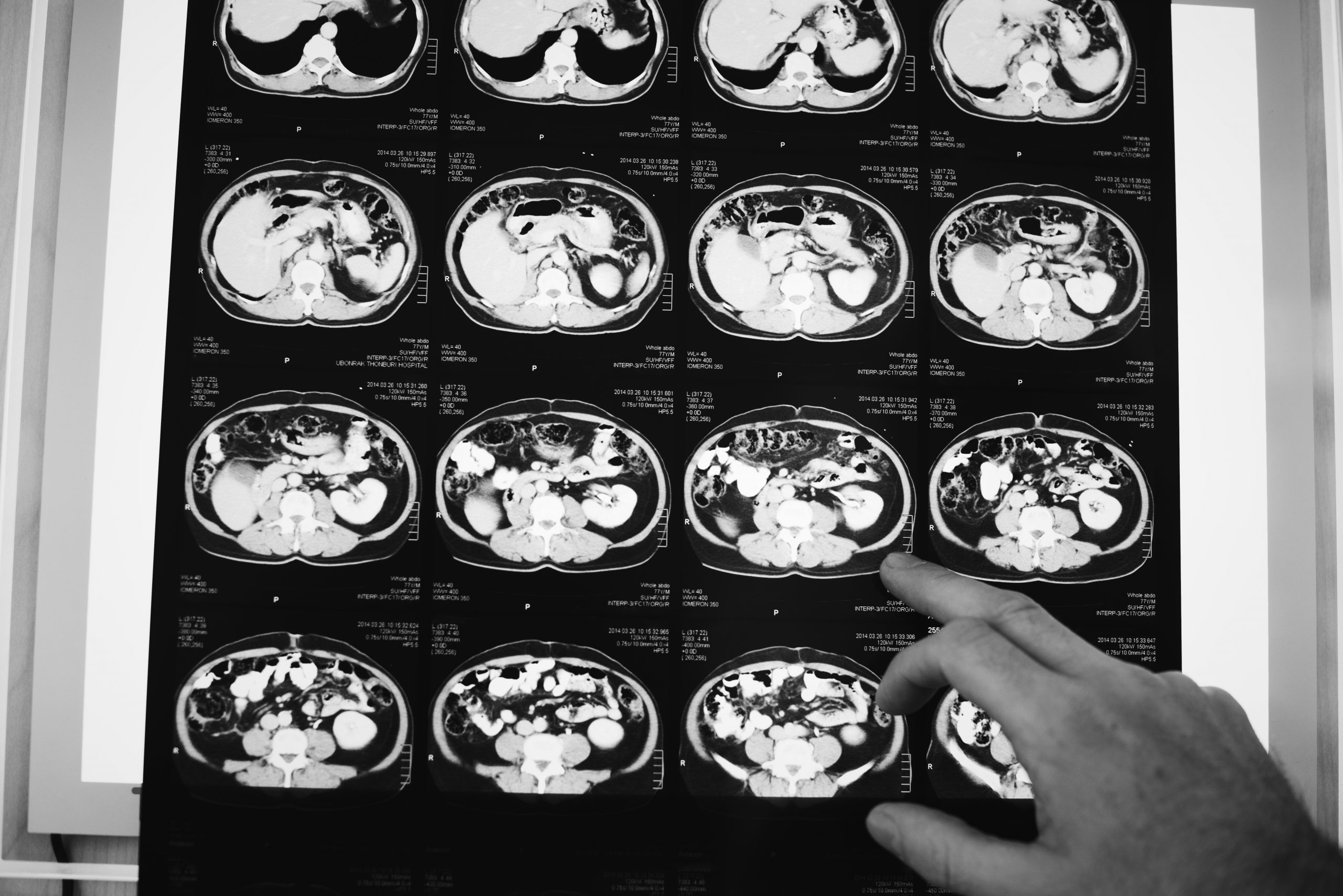
An abdominal magnetic resonance imaging scan is an imaging test that uses powerful magnets and radio waves. The waves create pictures of the inside of the belly area. It does not use radiation (x-rays).
Single magnetic resonance imaging (MRI) images are called slices. The images can be stored on a computer, viewed on a monitor, printed on film or scanned to a disk. One exam produces dozens or sometimes hundreds of images.

How the Test is Performed

You may be asked to wear a hospital gown or clothing without metal zippers or snaps (such as sweatpants and a t-shirt). Certain types of metal can cause blurry images.
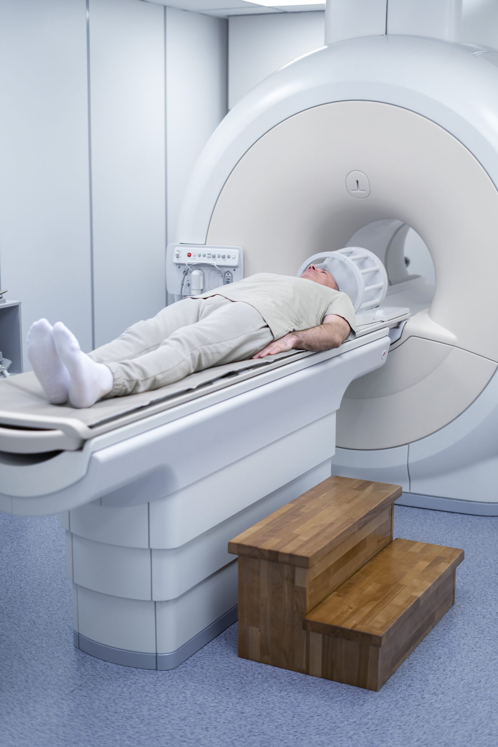
You will lie on a narrow table. The table slides into a large tunnel-shaped scanner.
Some exams require a special dye (contrast). Most of the time, the dye is given during the test through a vein (IV) in your hand or forearm. The dye helps the radiologist see certain areas more clearly.
During the MRI, the person who operates the machine will watch you from another room. The test lasts about 30 to 60 minutes, but it may take longer.

How to Prepare for the Test

You may be asked not to eat or drink anything for 4 to 6 hours before the scan.

Tell your health care provider if you are afraid of close spaces (have claustrophobia). You may be given a medicine to help you feel sleepy and less anxious. Your provider may also suggest an open MRI, in which the machine is not as close to your body.

Before the test, tell your provider if you have:

Because the MRI contains strong magnets, metal objects are not allowed into the room with the MRI scanner. Avoid carrying items such as:

How the Test will Feel

An MRI exam causes no pain. You may get medicine to relax you if you have a problem lying still or are very nervous. Moving too much can blur MRI images and cause errors.
The table may be hard or cold, but you can ask for a blanket or pillow. The machine makes loud thumping and humming noises when turned on. You can wear ear plugs to help reduce the noise.
An intercom in the room allows you to speak to someone at any time. Some MRI facilities have televisions and special headphones to help you pass time.
There is no recovery time, unless you were given a medicine to help you relax. After an MRI scan, you can go back to your normal diet, activity, and medicines.

Why the Test is Performed

An abdominal MRI provides detailed pictures of the belly area from many views. It is often used to clarify findings from earlier ultrasound or CT scan exams.

This test may be used to look at:

MRI can distinguish tumors from normal tissues. This can help your provider know more about the tumor such as size, severity, and spread. This is called staging.
In some cases it can give better information about masses in the abdomen than CT.

What Abnormal Results Mean

An abnormal result may be due to:

Risks

MRI does not use ionizing radiation. No side effects from the magnetic fields and radio waves have been reported.

The most common type of contrast (dye) used is gadolinium. It is very safe. Allergic reactions are rare but can occur. If you have a history of severe allergic reactions to other medicines you should notify your doctor. In addition, gadolinium can be harmful to people with kidney problems who need dialysis. Tell your provider before the test if you have kidney problems.

The strong magnetic fields created during an MRI can cause heart pacemakers and other implants not to work as well. The magnets can also cause a piece of metal inside your body to move or shift.

Alternative Names

  • Nuclear magnetic resonance – abdomen
  • NMR – abdomen
  • Magnetic resonance imaging – abdomen
  • MRI of the abdomen
  • Liver MRI – abdomen
  • Pancreas MRI – abdomen
  • Kidney MRI – abdomen

Lower leg

What is a lower leg MRI scan?

MRI stands for Magnetic Resonance Imaging, which is a way of taking pictures inside the body using magnetic fields and radio waves. It’s a safe, painless and non-invasive procedure capturing high-quality images. Unlike X-rays, MRI doesn’t use potentially harmful radiation.

The lower leg is comprised of bones, muscles, and other soft tissue including cartilage and nerves. A lower leg MRI scan produces detailed, cross-sectional images allowing your radiologist to examine the inner structure. 

What can a lower leg MRI scan detect?

The lower leg MRI scan will reveal any damage or abnormalities in your  

  • Bones (tibia and fibula)
  • Calf muscles (gastrocnemius and soleus)
  • Tendons and ligaments
  • Cartilage
  • Nerves

Depending on the results, your radiologist could diagnose any of a range of conditions, including 

  • Fractures
  • Soft tissue-related sprains
  • Muscle strains and tears
  • Nerve compression
  • Tendonitis
  • Osteoarthritis
  • Osgood-Schlatter disease
  • Achilles tendinopathy
  • Shin splints
  • Compartment syndrome

Armed with detailed knowledge of the cause of your lower leg pain, your physician will be able to plan and prescribe appropriate treatment. 

How do I prepare for a leg MRI scan?

Our patient care team will ensure you’re ready for your scan, but you can help by telling them about your medical history. Unless you’re asked to do otherwise, you can continue to eat, drink and take medication as normal before your scan.  

Some implants and conditions may affect how your MRI scan is performed, or whether it’s safe to do it at all. These include 

  • Cochlear implants
  • ICDs (implantable cardioverter-defibrillators)
  • Pacemakers
  • Artificial joints
  • Artificial heart valves
  • Allergy to MRI contrast material
  • Pregnancy
  • Claustrophobia

If any of these applies to you, please raise the issue with the patient care team before booking an appointment. 

What happens during a lower leg MRI scan, and how long does it take?

Your radiologist will ask you to take off any jewellery and empty your pockets, or you may be asked to change into a gown. You’ll then lie on the MRI table during the scan. A lower leg MRI scan takes around 40 minutes, and we would ask you to arrive around 15 minutes before your scheduled appointment. 

Arm

What is an arm MRI scan?

MRI stands for Magnetic Resonance Imaging, which is a way of taking pictures inside the body using magnetic fields and radio waves. It’s a safe, painless and non-invasive procedure capturing high-quality images. Unlike X-rays, MRI doesn’t use potentially harmful radiation.

An MRI scan will usually focus either on the upper arm between the elbow and shoulder, or the lower or forearm, depending on your symptoms and where you’re experiencing pain. 

What kinds of private arm MRI scans are available?

The arm is usually considered as two distinct parts, the upper arm and the lower or forearm. Your MRI scan will concentrate on one or the other, depending on what’s appropriate at the time. If it’s needed, you can schedule scans of each part of the arm together. 

Upper Arm 

An MRI scan will show images of your ligaments, tendons, humerus, and the four upper arm muscles – the biceps, brachialis, coracobrachialis and triceps. 

Lower or forearm 

The forearm is far more complex than the upper arm. It’s made up of two bones and 20 muscles. The MRI images will show the specific locations of any damage or abnormalities.

What symptoms would prompt an arm MRI scan?

If you’re experiencing any of the following symptoms, an arm MRI scan could diagnose the problem and help you plan treatment. 

  • Sharp or recurring pain
  • Limited mobility
  • Numbness
  • Inflammation or swelling

What can an arm MRI scan detect?

The results of your private MRI arm scan could help diagnose a range of injuries or conditions, including:

  • Bone fractures
  • Muscle or tendon tears
  • Infections
  • Nerve damage
  • Tumours
  • Osteoarthritis

An MRI scan is a diagnostic tool, so it won’t treat these conditions. However, the results will allow your physician to make informed decisions and recommend appropriate treatment. 

What happens during an arm MRI scan, and how long does it take?

Your radiologist will ask you to take off any jewellery and empty your pockets, or you may be asked to change into a gown. You’ll then lie on the MRI table during the scan. An arm MRI scan takes around 40 minutes, and we would ask you to arrive around 15 minutes before your scheduled appointment.

Neck (Soft tissue)

What is a neck soft tissue MRI scan?

MRI stands for Magnetic Resonance Imaging, which is a way of taking pictures inside the body using magnetic fields and radio waves. It’s a safe, painless and non-invasive procedure, capturing high-quality images. Unlike X-rays, MRI doesn’t use potentially harmful radiation. 
 
As the name suggests, a neck soft tissue MRI looks at the neck area, including the muscles, arteries and spinal cord, as well as the vertebrae and the lower part of the brain.  

What symptoms would prompt a neck soft tissue MRI scan?

If you’re experiencing severe neck pain that doesn’t appear to be getting better, it could be time for a neck soft tissue MRI scan. An MRI scan of the neck area could also be used before or after spinal surgery, to provide a guide for the operating surgeons.

How do I prepare for a neck MRI scan?

Our patient care team will ensure you’re ready for your scan, but you can help by telling them about your medical history. Unless you’re asked to do otherwise, you can continue to eat, drink and take medication as normal before your scan.  

Some implants and conditions may affect how your MRI scan is performed, or whether it’s safe to do it at all. These include 

  • Cochlear implants
  • ICDs (implantable cardioverter-defibrillators)
  • Pacemakers
  • Artificial joints
  • Artificial heart valves
  • Allergy to MRI contrast material
  • Pregnancy
  • Claustrophobia

If any of these applies to you, please raise the issue with the patient care team before booking an appointment. 

What happens during a neck soft tissue MRI scan, and how long does it take?

Your radiologist will ask you to take off any jewellery and empty your pockets, or you may be asked to change into a gown. You’ll then lie on the MRI table during the scan. A neck MRI scan takes around 40 minutes, and we would ask you to arrive around 15 minutes before your scheduled appointment.

Elbow

What is an elbow MRI scan?

MRI stands for Magnetic Resonance Imaging, which is a way of taking pictures inside the body using magnetic fields and radio waves. It’s a safe, painless and non-invasive procedure, which captures high-quality images. Unlike X-rays, MRI doesn’t use potentially harmful radiation.  

During an elbow MRI scan, the radiologist will use the scanner to take cross-section images of your elbow showing the structures within it, including  

  • Bones
  • Muscles
  • Tendons and ligaments
  • Cartilage
  • Other soft tissues

Why would I choose to have a private elbow MRI scan?

You should consider getting an MRI scan of your elbow if you’ve been experiencing problems that have lasted longer than a few weeks. Symptoms include 

  • Joint pain
  • Recurring muscle pain
  • Decreased mobility
  • Unexplained swelling
  • Sports injuries

What can an elbow MRI scan detect?

The elbow MRI scan will detect damage to tissues and bones, and abnormal growths. Some of the possible issues the scan can detect are 

  • Bone fractures
  • Ligament tears
  • Nerve damage
  • Inflammation
  • Dislocation
  • Osteoarthritis
  • Tumours

How do I prepare for an elbow MRI scan?

Our patient care team will ensure you’re ready for your scan, but you can help by telling them about your medical history. Unless you’re asked to do otherwise, you can continue to eat, drink and take medication as normal before your scan. 

Some implants and conditions may affect how your MRI scan is performed, or whether it’s safe to do it at all. These include 

  • Cochlear implants
  • ICDs (implantable cardioverter-defibrillators)
  • Pacemakers
  • Artificial joints
  • Artificial heart valves
  • Allergy to MRI contrast material
  • Pregnancy
  • Claustrophobia

If any of these applies to you, please raise the issue with the patient care team before booking an appointment. 

What happens during an elbow MRI scan, and how long does it take?

Your radiologist will ask you to take off any jewellery and empty your pockets, or you may be asked to change into a gown. You’ll then lie on the MRI table (or sit or stand in an upright MRI scanner) during the scan. An elbow MRI scan takes around 40 minutes, and we would ask you to arrive around 15 minutes before your scheduled

Calf

What is a calf MRI scan?

MRI stands for Magnetic Resonance Imaging, which is a way of taking pictures inside the body using magnetic fields and radio waves. It’s a safe, painless and non-invasive procedure capturing high-quality images. Unlike X-rays, MRI doesn’t use potentially harmful radiation.

The calf is comprised of bones, muscles, and other soft tissue including cartilage and nerves. A calf MRI scan produces detailed, cross-sectional images allowing your radiologist to examine the inner structure. 

What conditions might prompt me to get a calf MRI?

If you’re experiencing any of these symptoms, it could be time to get a calf MRI: 

  • Bone pain
  • Pain combined with fever
  • Instability of your foot or ankle
  • A lump or mass
  • Pain, discolouration, or inflammation

What can a calf MRI scan detect?

The calf MRI scan will reveal any damage or abnormalities in your  

  • Tibia
  • Fibula
  • Muscles
  • Tendons and ligaments
  • Nerves
  • Blood vessels
  • Additional soft tissues

Depending on the results, your radiologist could diagnose any of a range of conditions, including:

  • Muscle or ligament tears
  • Inflammation
  • Nerve damage
  • Infections

Armed with detailed knowledge of the cause of your calf pain, your physician will be able to plan and prescribe appropriate treatment.

How do I prepare for a calf MRI scan?

Our patient care team will ensure you’re ready for your scan, but you can help by telling them about your medical history. Unless you’re asked to do otherwise, you can continue to eat, drink, and take medication as normal before your scan.

Some implants and conditions may affect how your MRI scan is performed, or whether it’s safe to do it at all. These include:

  • Cochlear implants
  • ICDs (implantable cardioverter-defibrillators)
  • Pacemakers
  • Artificial joints
  • Artificial heart valves
  • Allergy to MRI contrast material
  • Pregnancy
  • Claustrophobia

If any of these applies to you, please raise the issue with the patient care team before booking an appointment.

What happens during a calf MRI scan, and how long does it take?

Your radiologist will ask you to take off any jewellery and empty your pockets, or you may be asked to change into a gown. You’ll then lie on the MRI table during the scan. A calf MRI scan takes around 20 minutes, and we would ask you to arrive around 15 minutes before your scheduled appointment.

Chest (Thorax)

What is a chest MRI scan?

MRI stands for Magnetic Resonance Imaging, which is a way of taking pictures inside the body using magnetic fields and radio waves. It’s a safe, painless, and non-invasive procedure capturing high-quality images. Unlike X-rays, MRI doesn’t use potentially harmful radiation.

A chest (thorax) MRI scan will produce detailed, cross-sectional images of the inside of your chest cavity. It’s important to note that this procedure does not examine the heart – if you’re concerned about heart health, please see details of our cardiac MRI scans here.

Fast, affordable private MRI scans for the chest (thorax)

The chest is a hard-working part of the body, and long-term pain or discomfort here can have a significant impact on your overall quality of life. An MRI scan of your chest will provide detailed images of the chest cavity, including the mediastinum, chest wall, pleura, and blood vessels.

What symptoms would prompt a private chest MRI scan?

If you’re experiencing severe or long-term symptoms, or if the cause is difficult to pinpoint using other diagnostic tools, a chest MRI may be the best way forward. Symptoms that might prompt a scan include sharp pain when breathing, or a constant tightness in your chest.

What can a chest MRI scan detect?

Your private chest MRI scan will take detailed, cross-sectional images of the inside of your chest cavity. The body parts you’ll see in these images include:

  • Thymus gland
  • Oesophagus
  • Trachea
  • Thoracic diaphragm
  • Other soft tissues
  • Chest wall (skin, fat, muscles, and bones)

By examining the images, your radiologist will be able to determine if there is any damage or abnormal growths present. You may be diagnosed with any of the following conditions:

  • Tumours (including size and degree of spread)
  • Organ diseases
  • Injuries

With a detailed diagnostic report, your physician will be able to determine the best course of action for your treatment.

How do I prepare for a chest MRI scan?

Our Patient Care Team will ensure you’re ready for your scan, but you can help by telling them about your medical history. Unless you’re asked to do otherwise, you can continue to eat, drink, and take medication as normal before your scan.

Some implants and conditions may affect how your MRI scan is performed, or whether it’s safe to do it at all. These include:

  • Cochlear implants
  • ICDs (implantable cardioverter-defibrillators)
  • Pacemakers
  • Artificial joints
  • Artificial heart valves
  • Allergy to MRI contrast material
  • Pregnancy
  • Claustrophobia

If any of these applies to you, please raise the issue with the patient care team before booking an appointment.

What happens during a chest or thorax MRI scan, and how long does it take?

Your radiologist will ask you to take off any jewellery and empty your pockets, or you may be asked to change into a gown. You’ll then lie on the MRI table during the scan. A chest MRI scan takes around 40 minutes, and we would ask you to arrive around 15 minutes before your scheduled appointment.

Foot & Ankle

Get your foot and ankle problems diagnosed quickly

Feet and ankles are fundamental to your mobility. This is a complex part of the body – with each foot consisting of 26 bones and 30 joints – making it difficult to pinpoint the cause of pain. A foot or ankle MRI scan can identify and diagnose the injury or condition that’s causing the problem.

Please note that the foot and ankle are separate scans.

What is a foot or ankle MRI scan?

MRI stands for Magnetic Resonance Imaging, which is a way of taking pictures inside the body using magnetic fields and radio waves. It’s a safe, painless and non-invasive procedure, which captures high-quality images. Unlike X-rays, MRI doesn’t use potentially harmful radiation.

The area around the foot and ankle is complex – there are 26 bones, 30 joints and 29 muscles in the foot alone. Depending on your symptoms and where you’re experiencing pain, your radiologist will scan your forefoot, midfoot or hindfoot, your ankle, or your Achilles tendon.

Each of these areas is composed of different bones, as in this list:

Forefoot :

  • Metatarsal bones
  • Proximal phalanges
  • Middle phalanges
  • Distal phalanges

Midfoot :

  • Navicular bone
  • Cuboid bone
  • Three cuneiform bones

Hindfoot :

  • Talus bone
  • Calcaneus bone

Ankle :

  • Talus bone
  • Bottom part of the tibia
  • Bottom part of the fibula
  • Connection to the talus bone

Achilles tendon:

An Achilles tendon MRI scan will show the condition of the tendon connecting your calf muscle to your hindfoot.

Why would I choose to have a private foot or ankle MRI scan?

If you’re experiencing any of these symptoms, you may want to arrange a private foot or ankle MRI scan:

  • Pain
  • Swelling
  • Stiffness
  • Loss of mobility
  • Bruising
  • An inability to put weight on your foot without pain

What can a foot or ankle MRI scan detect?

Your foot or ankle MRI scan can detect and assess a wide range of injuries and conditions, depending on which area is being scanned. Among other conditions, your scan will be looking for:

Forefoot:

  • Fractures
  • Claw toe
  • Mallet toe
  • Bunions
  • Gout (most often affects the big toe)

Midfoot:

  • Fractures
  • Bursitis
  • Inflammation

Hindfoot:

  • Fractures
  • Heel spur
  • Plantar fasciitis
  • Bursitis

Ankle:

  • Fractures
  • Ligament sprain (ankle sprains are the most common, but they can happen in any other part)

Achilles tendon:

  • Achilles tendon rupture
  • Achilles tendinitis

How do I prepare for a foot and ankle MRI scan?

Our patient care team will ensure you’re ready for your scan, but you can help by telling them about your medical history. Unless you’re asked to do otherwise, you can continue to eat, drink and take medication as normal before your scan.

Some implants and conditions may affect how your MRI scan is performed, or whether it’s safe to do it at all. These include:

  • Cochlear implants
  • ICDs (implantable cardioverter-defibrillators)
  • Pacemakers
  • Artificial joints
  • Artificial heart valves
  • Allergy to MRI contrast material
  • Pregnancy
  • Claustrophobia

If any of these applies to you, please raise the issue with the patient care team before booking an appointment.

What happens during a foot or ankle MRI scan, and how long does it take?

Your radiologist will ask you to take off any jewellery and empty your pockets, or you may be asked to change into a gown. You’ll then lie on the MRI table during the scan. A foot or ankle MRI scan takes around 20 minutes, and we would ask you to arrive around 15 minutes before your scheduled appointment.

Thigh

What is a thigh MRI scan?

MRI stands for Magnetic Resonance Imaging, which is a way of taking pictures inside the body using magnetic fields and radio waves. It’s a safe, painless, and non-invasive procedure capturing high-quality images. Unlike X-rays, MRI doesn’t use potentially harmful radiation.

The thigh is comprised of bones, muscles, and other soft tissue including cartilage and nerves. A thigh MRI scan produces detailed, cross-sectional images allowing your radiologist to examine the inner structure.

What can a thigh MRI scan detect?

The thigh MRI scan will reveal any damage or abnormalities in your:

  • Muscles
  • Bones
  • Tendons and ligaments
  • Cartilage
  • Other soft tissues

Depending on the results, your radiologist could diagnose any of a range of conditions, including:

  • Fractures
  • Ligament tears
  • Muscle or tendon strain
  • Bone tumours
  • Compartment syndrome
  • Iliotibial band syndrome

Armed with detailed knowledge of the cause of your upper leg pain, your physician will be able to plan and prescribe appropriate treatment.

How do I prepare for a thigh MRI scan?

Our patient care team will ensure you’re ready for your scan, but you can help by telling them about your medical history. Unless you’re asked to do otherwise, you can continue to eat, drink, and take medication as normal before your scan.

Some implants and conditions may affect how your MRI scan is performed, or whether it’s safe to do it at all. These include:

  • Cochlear implants
  • ICDs (implantable cardioverter-defibrillators)
  • Pacemakers
  • Artificial joints
  • Artificial heart valves
  • Allergy to MRI contrast material
  • Pregnancy
  • Claustrophobia

If any of these applies to you, please raise the issue with the patient care team before booking an appointment.

What happens during a thigh MRI scan, and how long does it take?

Your radiologist will ask you to take off any jewellery and empty your pockets, or you may be asked to change into a gown. You’ll then lie on the MRI table during the scan. An upper leg MRI scan takes around 20 minutes, and we would ask you to arrive around 15 minutes before your scheduled appointment.

Shoulder

What is a shoulder MRI scan?

MRI stands for Magnetic Resonance Imaging, which is a way of taking pictures inside the body using magnetic fields and radio waves. It’s a safe, painless, and non-invasive procedure that captures high-quality images. Unlike X-rays, MRI doesn’t use potentially harmful radiation.

During a shoulder MRI scan, the radiologist will use the scanner to take cross-sectional images of your shoulder showing the structures within it, including:

  • Bones (clavicle, head of the humerus)
  • Muscles (deltoid and rotator cuff)
  • Tendons and ligaments
  • Cartilage
  • Nerves
  • Other soft tissues

Why would I choose to have a private shoulder MRI scan?

You should consider getting an MRI scan of your shoulder if you’ve been experiencing problems that have lasted longer than a few weeks. Symptoms include:

  • Joint pain
  • Recurring muscle pain
  • Decreased mobility
  • Unexplained swelling
  • Sports injuries

What can a shoulder MRI scan detect?

The shoulder MRI scan will detect damage to tissues and bones, as well as abnormal growths. Some of the possible issues the scan can detect are:

  • Arthritis, labral tears, and other degenerative joint disorders
  • Fractures
  • Rotator cuff disorders
  • Ligament or tendon tears
  • Infections
  • Swelling or bleeding in the tissues

How do I prepare for a shoulder MRI scan?

Our Patient Care Team will ensure you’re ready for your scan, but you can help by telling them about your medical history. Unless you’re asked to do otherwise, you can continue to eat, drink, and take medication as normal before your scan.

Some implants and conditions may affect how your MRI scan is performed, or whether it’s safe to do it at all. These include:

  • Cochlear implants
  • ICDs (implantable cardioverter-defibrillators)
  • Pacemakers
  • Artificial joints
  • Artificial heart valves
  • Allergy to MRI contrast material
  • Pregnancy
  • Claustrophobia

If any of these applies to you, please raise the issue with the patient care team before booking an appointment.

What happens during a shoulder MRI scan, and how long does it take?

Your radiologist will ask you to take off any jewellery and empty your pockets, or you may be asked to change into a gown. You’ll then lie on the MRI table during the scan. A shoulder MRI scan takes around 20 minutes, and we would ask you to arrive around 15 minutes before your scheduled appointment.

Cardiac (Heart)

What is a cardiac MRI scan?

MRI stands for magnetic resonance imaging. It allows you to take pictures of your body by using a strong magnetic field and radio frequency pulses. A cardiac MRI scan produces images of the heart muscle and valves and is used to assess both its structure and function.

A cardiac MRI is used to diagnose a wide range of heart conditions, including congenital heart disease, heart valve disease, and cardiac tumors. MRI scanning is widely performed, and unlike X-ray, it does not use ionizing radiation and therefore is not harmful.

What can a cardiac MRI scan show?

  • Assess the structure and function of your heart chambers, valves, and major blood vessels.
  • Diagnose cardiovascular problems such as cancers, tumors, and inflammatory conditions.
  • Detect the blockage of blood vessels.

Image slices are taken during the scan while the MRI moves across three planes:

  • Sagittal – From side to side
  • Axial – From top to bottom
  • Coronal – From front to back

What can a cardiac MRI scan diagnose?

A cardiac MRI scan can diagnose a variety of conditions related to the heart, including:

  • Heart valve abnormalities
  • Congenital heart disease (CHD) or congenital heart defects
  • Cardiomyopathy (enlarged or thickened heart muscle)
  • Cardiac tumors or masses in the heart

Direct insight into your heart

Get full insight into the inside of your heart. Receive an expert diagnosis and evaluation of any cardiac abnormality, including those involving your heart muscle.

Pelvis

What is a pelvis or groin MRI scan?

MRI stands for Magnetic Resonance Imaging, which is a way of taking pictures inside the body using magnetic fields and radio waves. It’s a safe, painless and non-invasive procedure capturing high-quality images. Unlike X-rays, MRI doesn’t use potentially harmful radiation.

The pelvic area includes a diverse group of organs, and a wide range of symptoms could prompt an MRI scan. A thorough clinical history is always helpful in deciding on the correct scan examination

What kind of MRI pelvis and groin scans are available?

We offer different types of pelvis MRI scans:

  • Pelvis Musculoskeletal MRI
  • Female Pelvis MRI Scan (Gynaecological)
  • Sacroiliac Joints Scan
  • Groin scan
  • Prostate scan

We also offer a dedicated, specialized prostate screening service. See details here.

What symptoms would prompt a pelvis or groin MRI scan?

If you’re experiencing any of the following symptoms, it may be a good idea to get a pelvic MRI scan:

  • Pelvic pain
  • Hip pain
  • Stiffness or loss of mobility
  • Inflammation

What can a pelvis MRI scan detect?

We offer different pelvic MRI scans, focused on specific areas and conditions:

Pelvis Musculoskeletal

A scan of your pelvis will capture images of your: Pelvic girdle Muscles and ligaments Other soft tissues Any damage or abnormalities in these parts will show up in the images. You may be diagnosed with any of these conditions: Fractures Bone bruises Ligament and tendon tears Muscle strains Osteoarthritis Bursitis Infections Dislocation Tumours

Pelvis Gynaecological MRI scan

This looks at the female reproductive organs and associated organs, namely the uterus, ovaries, bladder and rectum.

Prostate MRI scan

For men, this scan examines the bladder, rectum and prostate gland. A specialist prostate screening service is also available.

Sacroiliac Joints Scan

This will show the sacrum, the ilium, and the surrounding tissues at the base of the spine. The images may indicate the presence of sacroiliitis, which is an inflammation of one or both joints, or sacroiliac joint dysfunction.


Groin Scan

The groin is the creased area joining the thigh to the torso, on either side of the pubic bone. The main reasons for assessing the groin are signs of muscle strain or inguinal hernia.

How do I prepare for a pelvis MRI scan?

Our patient care team will ensure you’re ready for your scan, but you can help by telling them about your medical history. Unless you’re asked to do otherwise, you can continue to eat, drink and take medication as normal before your scan.  

Some implants and conditions may affect how your MRI scan is performed, or whether it’s safe to do it at all. These include 

  • Cochlear implants
  • ICDs (implantable cardioverter-defibrillators)
  • Pacemakers
  • Artificial joints
  • Artificial heart valves
  • Allergy to MRI contrast material
  • Pregnancy
  • Claustrophobia

If any of these applies to you, please raise the issue with the patient care team before booking an appointment.

What happens during a pelvis MRI scan, and how long does it take?

Your radiologist will ask you to take off any jewellery and empty your pockets, or you may be asked to change into a gown. You’ll then lie on the MRI table during the scan. A pelvic MRI scan takes around 20-40 minutes, and we would ask you to arrive around 15 minutes before your scheduled appointment.

Mouth & Jaw

What is a mouth and jaw (TMJ) MRI scan?

MRI stands for Magnetic Resonance Imaging, which is a way of taking pictures inside the body using magnetic fields and radio waves. It’s a safe, painless, and non-invasive procedure, capturing high-quality images. Unlike X-rays, MRI doesn’t use potentially harmful radiation.

A TMJ MRI scan will take cross-sectional images of the inside of your mouth and jaw to examine the jaw muscles, the mandible and teeth, and the associated cartilage discs.

What symptoms would prompt a TMJ MRI scan?

Typical symptoms which might warrant a mouth and jaw TMJ MRI scan include:

  • Pain that travels through your jaw, face, and neck
  • Pain in your jaw or near your ear
  • Difficulty swallowing or speaking
  • Stiff jaw muscles
  • Loose teeth
  • Painful clicking or popping in the jaw

A doctor might also advise a mouth and jaw MRI scan if it’s difficult to determine the reasons for your symptoms or to gather more information to develop a treatment plan.

What can a mouth and jaw MRI scan detect?

During your scan, the radiologist will take cross-sectional images of the inside of your mouth and jaw using an MRI scanner. These will be used to examine your five jaw muscles, your mandible and teeth, and cartilage discs.

Your scan could reveal any of these conditions:

  • Dislocation
  • Fractures
  • Temporomandibular joint dysfunction
  • Osteonecrosis
  • Odontogenic tumours
  • Injuries to bones or tissues

You’ll then be able to take your report to an oral pathologist, who will use the information to determine the best treatment for your condition.

How do I prepare for a TMJ MRI scan?

Our patient care team will ensure you’re ready for your scan, but you can help by telling them about your medical history. Unless you’re asked to do otherwise, you can continue to eat, drink, and take medication as normal before your scan.

Some implants and conditions may affect how your MRI scan is performed, or whether it’s safe to do it at all. These include:

  • Cochlear implants
  • ICDs (implantable cardioverter-defibrillators)
  • Pacemakers
  • Artificial joints
  • Artificial heart valves
  • Allergy to MRI contrast material
  • Pregnancy
  • Claustrophobia

If any of these applies to you, please raise the issue with the patient care team before booking an appointment.

What happens during a mouth and jaw MRI scan, and how long does it take?

Your radiologist will ask you to take off any jewellery and empty your pockets, or you may be asked to change into a gown. You’ll then lie on the MRI table during the scan. A mouth and jaw MRI scan takes around 40 minutes, and we would ask you to arrive around 15 minutes before your scheduled appointment.

Small Bowel

What is small bowel or small intestine MRI scan?

MRI stands for Magnetic Resonance Imaging, which is a way of taking pictures inside the body using magnetic fields and radio waves. It’s a safe, painless, and non-invasive procedure capturing high-quality images. Unlike X-rays, MRI doesn’t use potentially harmful radiation.

An MRI scan of the small bowel is often recommended to investigate symptoms like chronic diarrhoea or constipation and may be used to monitor progress when treatment is underway.

What symptoms would prompt a private small bowel MRI scan?

If you’re experiencing any of the following symptoms, with no obvious causes, it may be a good idea to get a small bowel MRI scan:

  • Diarrhoea
  • Constipation
  • Gas
  • Vomiting
  • Swollen, painful belly
  • Blood in your stool
  • Sudden weight loss

What can a small bowel MRI scan detect?

Your small intestine MRI scan will show all three parts of the small bowel – the duodenum, the jejunum, and the ileum. It will identify any damage or abnormal growths. Typical conditions which could be diagnosed include:

  • Irritable bowel syndrome
  • Functional gastrointestinal disorders
  • Antibiotic-associated diarrhoea
  • Obscure bleeding
  • Malabsorption (difficulty digesting or absorbing nutrients)
  • Crohn’s disease of the ileum
  • Regional enteritis
  • Iron deficiency anaemia
  • Celiac disease
  • Gluten disorders

Once your radiologist identifies the cause of your symptoms, you can take the diagnostic report to a doctor, who will use it to prescribe an effective treatment.

How do I prepare for a small bowel MRI scan?

Our patient care team will ensure you’re ready for your scan, but you can help by telling them about your medical history. Unless you’re asked to do otherwise, you can continue to eat, drink, and take medication as normal before your scan.

Some implants and conditions may affect how your MRI scan is performed, or whether it’s safe to do it at all. These include:

  • Cochlear implants
  • ICDs (implantable cardioverter-defibrillators)
  • Pacemakers
  • Artificial joints
  • Artificial heart valves
  • Allergy to MRI contrast material
  • Pregnancy
  • Claustrophobia

If any of these apply to you, please raise the issue with the patient care team before booking an appointment.

What happens during a lower bowel MRI scan, and how long does it take?

Your radiologist will ask you to take off any jewellery and empty your pockets, or you may be asked to change into a gown. You’ll then lie on the MRI table during the scan. A lower intestine MRI scan takes around 60 minutes, and we would ask you to arrive around 15 minutes before your scheduled appointment.

Similar Services