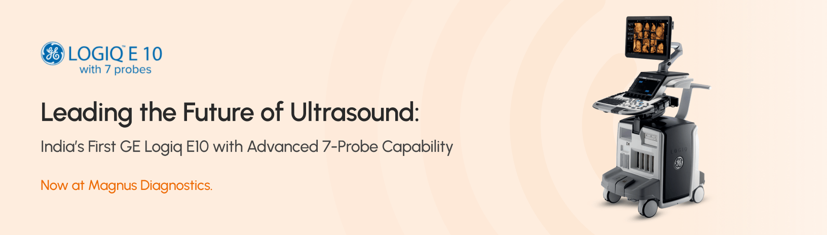
Magnus Diagnostics was founded with a commitment to redefine healthcare excellence by delivering world-class diagnostic services to our community and beyond.
Home > Our Service > Expert Ultrasound Scanning Services in Thrissur
What is an ultrasound?
Ultrasound (also called sonography or ultrasonography) is a non-invasive imaging test. An ultrasound picture is called a sonogram. Ultrasound uses high-frequency sound waves to create real-time pictures or video of internal organs or other soft tissues, such as blood vessels.
Ultrasound enables healthcare providers to “see” details of soft tissues inside your body without making any incisions (cuts). And unlike X-rays, ultrasound doesn’t use radiation. Although most people associate ultrasound with pregnancy, healthcare providers use ultrasound for many different situations and to look at several different parts of the inside of your body.
How does an ultrasound work?
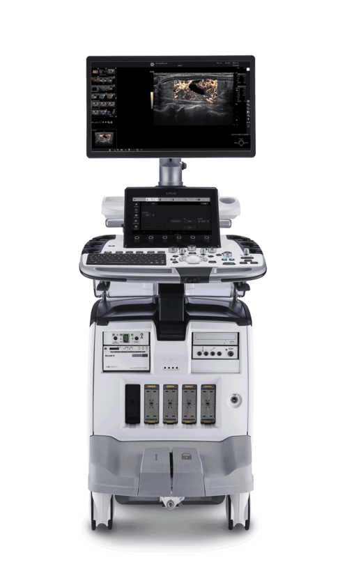
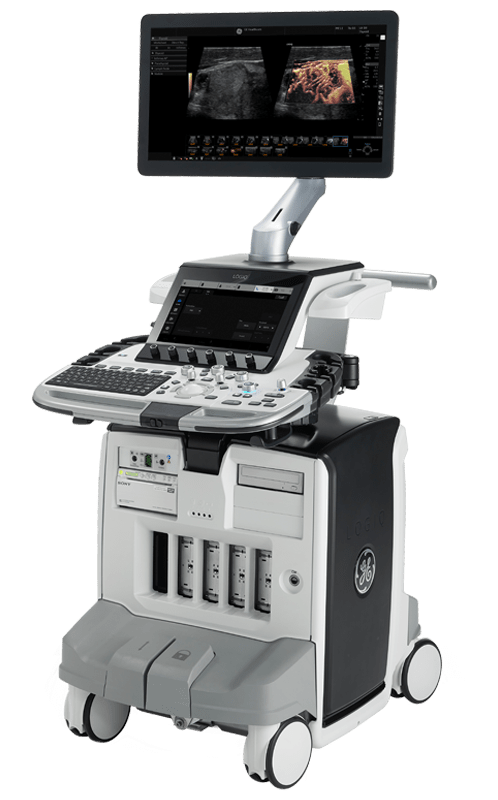
During an ultrasound, a healthcare provider passes a device called a transducer or probe over an area of your body or inside a body opening. The provider applies a thin layer of gel to your skin so that the ultrasound waves are transmitted from the transducer through the gel and into your body.
The probe converts electrical current into high-frequency sound waves and sends the waves into your body’s tissue. You can’t hear the sound waves. Sound waves bounce from structures inside your body and back to the probe, which converts the waves into electrical signals. A computer then converts the pattern of electrical signals into real-time images or videos, which are displayed on a computer screen nearby.
What are the different kinds of ultrasounds?
There are three main categories of ultrasound imaging, including:
Pregnancy ultrasound (prenatal ultrasound).
Diagnostic ultrasound.
Ultrasound guidance for procedures.
Pregnancy ultrasound
Healthcare providers often use ultrasound (often called prenatal or obstetric ultrasound) to monitor you and the foetus during pregnancy.
Providers use prenatal ultrasound to:
Confirm that you’re pregnant.
Check to see if you’re pregnant with more than one foetus.
Estimate how long you’ve been pregnant and the gestational age of the foetus.
Check the fetal growth and position.
See the foetal movement and heart rate.
Check for congenital conditions (birth defects) in the foetal brain, spinal cord, heart or other parts of its body.
Check the amount of amniotic fluid.
Most healthcare providers recommend an ultrasound at 20 weeks pregnant. This test tracks the foetus’s growth and development during pregnancy. This ultrasound may also show the biological sex of the foetus. Tell your technician if you do or do not want to know the sex.
Your provider may order extra scans to get answers to any questions or concerns, such as the potential for congenital conditions.
Diagnostic ultrasound
Providers use diagnostic ultrasounds to view internal parts of your body to see if something is wrong or not working properly. They can help your provider learn more about what’s causing a wide range of symptoms, such as unexplained pain, masses (lumps) or what may be causing an abnormal blood test. For most diagnostic ultrasound exams, the technician places the transducer (probe) on your skin. In some cases, they may need to place the probe inside your body, such as in your vagina or rectum.
The type of diagnostic ultrasound you have depends on the details of your case. Examples of diagnostic ultrasounds include:
Abdominal ultrasound
An ultrasound probe moves across the skin of your midsection (belly) area. Abdominal ultrasound can diagnose many causes of abdominal pain.
Kidney (renal) ultrasound
Providers use kidney ultrasound to assess the size, location and shape of your kidneys and related structures, such as your ureters and bladder. Ultrasound can detect cysts, tumours, obstructions or infections within or around your kidneys.
Breast ultrasound
A breast ultrasound is a non-invasive test to identify breast lumps and cysts. Your provider may recommend an ultrasound after an abnormal mammogram.
Doppler ultrasound
This is a special ultrasound technique that assesses the movement of materials, like blood, in your body. It allows your provider to see and evaluate blood flow through arteries and veins in your body. Doppler ultrasound is often used as part of a diagnostic ultrasound study or as part of a vascular ultrasound.
Pelvic ultrasound
A pelvic ultrasound looks at the organs in your pelvic area between your lower abdomen (belly) and legs. Some of the pelvic organs include your bladder, prostate, rectum, ovaries, uterus and vagina.
Transvaginal ultrasound
Your provider inserts a probe into your vaginal canal. It shows reproductive tissues such as your uterus or ovaries. A transvaginal ultrasound is sometimes called a pelvic ultrasound because it evaluates structures inside your pelvis (hip bones).
Thyroid ultrasound
Providers use ultrasound to assess your thyroid, a butterfly-shaped endocrine gland in your neck. Providers can measure the size of your thyroid and see if there are nodules or lesions within the gland.
Transrectal ultrasound
Your provider inserts an ultrasound probe transducer into your rectum. It evaluates your rectum or other nearby tissues, such as the prostate in people assigned male at birth.
Ultrasound guidance for procedures
Providers sometimes use ultrasound to perform certain procedures precisely. A common use of ultrasound is to guide needle placement to sample fluid or tissue from:
Tendons
Joints
Muscles
Cysts or fluid collections
Soft-tissue masses
Organs (liver, kidney or prostate)
Transplant organs (liver, kidney or pancreas)
Examples of other procedures that may require ultrasound guidance include:
Embryo transfer for in vitro fertilization
Nerve blocks
Confirming the placement of an IUD (intrauterine device) after insertion
Lesion localization procedures
What is the difference between a 3D ultrasound and a 4D ultrasound?
For ultrasounds during pregnancy, the traditional ultrasound is a two-dimensional (2D) image of the foetus. 2D ultrasound produces outlines and flat looking images, which allows your healthcare provider to see the foetus’s internal organs and structures.
Three-dimensional (3D) ultrasound allows the visualization of some facial features of the foetus and possibly other body parts such as fingers and toes. Four-dimensional (4D) ultrasound is 3D ultrasound in motion. Providers rarely use 3D or 4D foetal ultrasound imaging for medical purposes, though it can be useful in diagnosing a facial or skeletal issue. They do, however, use 3D ultrasound for other medical purposes, such as evaluating uterine polyps and fibroids.
While ultrasound is generally considered to be safe with very low risks, the risks may increase with unnecessary prolonged exposure to ultrasound energy or when untrained users operate an ultrasound machine. Because of this, the U.S. Food and Drug Administration (FDA) advises against getting a 3D ultrasound for non-medical reasons such as for “keepsake” moments or entertainment.
Who performs an ultrasound?
A doctor or a healthcare provider called an ultrasound technician or sonographer performs ultrasounds. They’re specially trained to operate an ultrasound machine properly and safely. It’s important to always have your ultrasound performed by a medical professional and in a medical facility.
What happens during an ultrasound?
Preparation for an ultrasound varies depending on what body part you’ll have scanned. Your provider may ask you to remove certain pieces of clothes or change into a hospital gown.
Ultrasounds that involve applying the transducer (probe) over your skin (not in your body), follow these general steps:
You’ll lie on your side or back on a comfortable table
The ultrasound technician will apply a small amount of water-soluble gel on your skin over the area to be examined. This gel doesn’t harm your skin or stain your clothes
The technician will move a handheld transducer or probe over the gel to get images inside your body
The technician may ask you to be very still or to hold your breath for a few seconds to create clearer pictures
Once the technician has gotten enough images, they’ll wipe off any remaining gel on your skin, and you’ll be done
An ultrasound test usually takes 30 minutes to an hour. If you have any questions about your specific type of ultrasound, ask your healthcare provider.
Are ultrasounds safe in pregnant women?
Yes, ultrasound is considered safe during pregnancy. It uses sound waves, not radiation, to create images of the developing baby, uterus, and surrounding tissues. Numerous studies and years of clinical use have shown no known risks to the mother or fetus when ultrasounds are used appropriately.
Ultrasound during pregnancy is a valuable tool that can:
Monitor fetal development and growth
Detect certain birth defects or abnormalities
Check the position of the fetus and placenta
Measure amniotic fluid levels
Most healthcare providers recommend ultrasounds at key stages in pregnancy, such as the early pregnancy scan (around 6-10 weeks) and the detailed anomaly scan (around 18-22 weeks). When used for medical purposes and by trained professionals, ultrasound is a safe and effective way to support prenatal care.
What conditions can be detected by ultrasound?
Ultrasound can help providers diagnose a wide range of medical issues, including:
Abnormal growths, such as tumours or cancer
Blood clots
Enlarged spleen
Ectopic pregnancy (when a fertilized egg implants outside of your uterus)