CT Scan, MRI, or X-Ray: Which One Do You Need?
Diagnostic imaging has become an essential part of modern healthcare, helping doctors detect, diagnose, and treat a wide range of conditions. From identifying a simple fracture to locating internal bleeding or evaluating complex issues in the brain and spinal cord, these tests provide accurate insights without invasive procedures. In Thrissur, where advanced medical facilities are steadily growing, patients often have access to X-rays, CT scans, and MRI services, but choosing the right test can still be confusing.
Understanding the key differences between an X-ray, CT scan, and MRI is essential for every patient to get a clear pathway in their healthcare journey. This guide will take you through each imaging test, explaining how each works, what conditions they are best suited for, and how they differ from each other.
What is a CT Scan?
A CT (Computed Tomography) scan is an advanced imaging procedure that utilises specialised X-ray techniques and computer technology to generate pictures of the internal organs, bones, and other structures of the body.
As a non-invasive and painless procedure, CT scans are often preferred to detect a variety of medical conditions. Unlike a regular X-ray, which provides a flat, two-dimensional image, a CT scan is more advanced and can show bones, blood vessels, and soft tissues in greater detail.
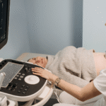
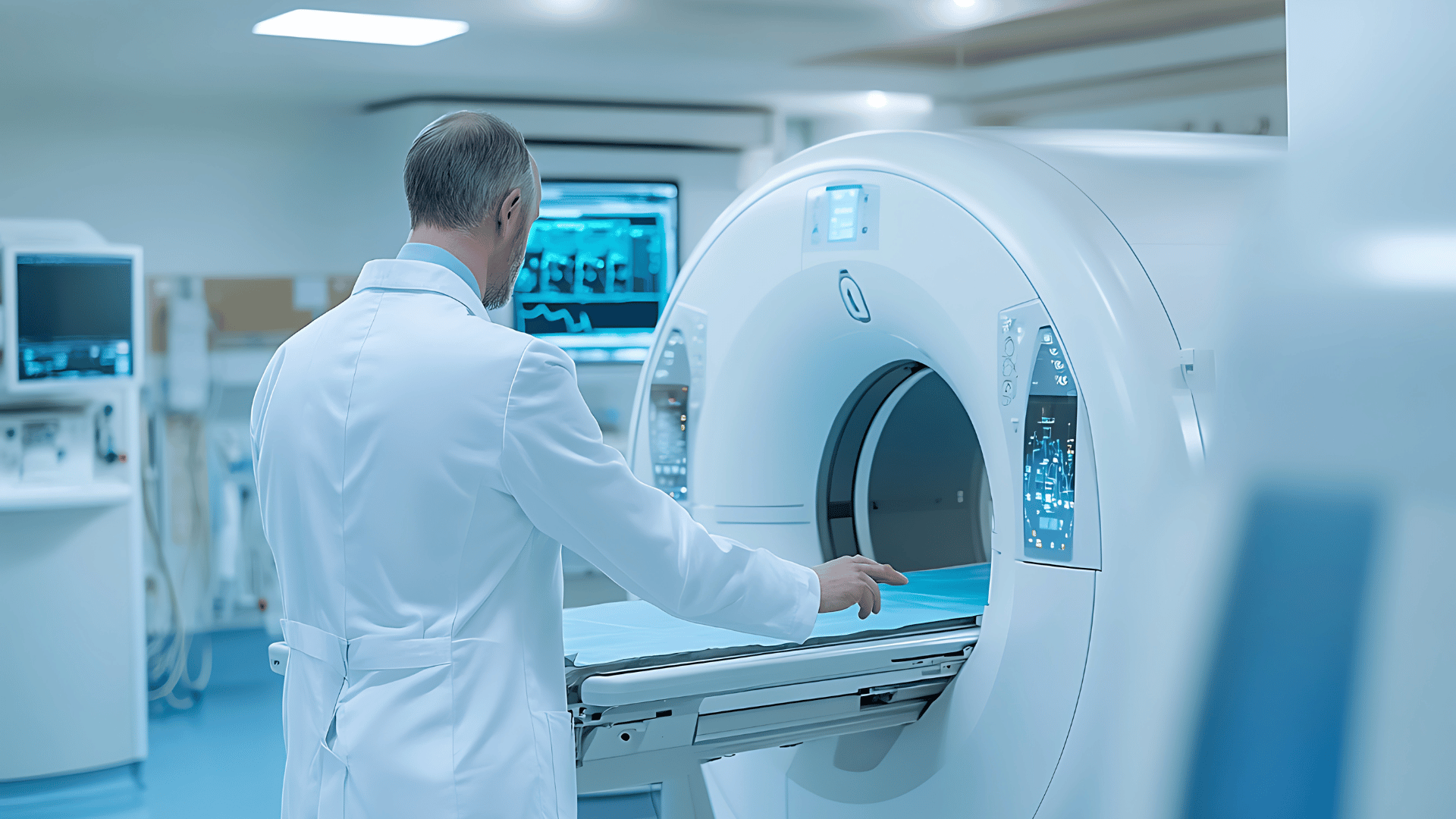
During a CT scan, the patient lies on a table that slowly moves through a large, circular machine. The X-ray beams rotate around the body and capture multiple images from different angles. These images are then processed by a computer to generate highly detailed sections of the area being examined.
Uses of CT Scans
A CT scan is one of the most commonly used and versatile medical tests and is used for:
- Diagnosing and monitoring diseases, from minor infections to life-threatening conditions like cancer.
- Assessing bone injuries and conditions, including fractures or tumours.
- Guiding certain procedures, such as biopsies or surgeries.
- Monitoring the effectiveness of treatments.
- Detecting internal injuries or bleeding after trauma.
Types of CT Scan
CT scans exist in different types and are used depending on the medical condition:
1. Positron Emission Tomography (PET) CT Scan
A Positron Emission Tomography (PET) scan makes use of radioactive materials to diagnose diverse conditions, including tumours, heart disease, or brain disorders. It is a hybrid imaging technique that combines positron emission tomography (PET) and traditional CT.
This scan is highly effective for detecting cancers, monitoring response to treatment, and identifying areas of abnormal cell activity. The combination of PET and CT improves diagnostic accuracy, especially for tumours and active disease sites that may not be visible on standard CT alone.
2. CT Urography
CT urography is a specialised CT scan focused on the urinary tract, including the kidneys, bladder, and ureters. A contrast dye is often used to highlight abnormalities such as kidney stones, tumours, or urinary blockages.
The contrast dye is injected into the vein, which outlines the urinary system to produce detailed cross-sectional images. Images are captured in multiple phases, including non-contrast, nephrographic, and excretory phases, to highlight different parts of the urinary tract. This multiphase approach improves the ability to detect and characterise a wide range of abnormalities, such as stones, tumours, and structural defects.
What is an MRI?
An MRI (Magnetic Resonance Imaging) scan is a type of scan that uses strong magnetic fields and radio waves to create images of the inside of the body, including organs, tissues, and other internal structures. It is often the most preferred diagnostic tool for evaluating soft tissues such as the brain, spinal cord, joints, and internal organs.
During an MRI, the patient lies in a tunnel-like machine while the scanner creates strong magnetic fields. These fields cause hydrogen atoms in the body to align temporarily, and radio waves then generate signals that the computer translates into images.
Types of MRI Scan
There are several types of MRI scans, each used for diverse diagnostic purposes. Here are some of the different types of MRI scans:
-
Open MRI Scans
Open MRI scans are specially designed to ensure patient comfort by reducing claustrophobia and movement restrictions. These scanners feature an open design with fewer enclosure walls. They function on relatively lower magnetic field strengths; therefore, the image quality and resolution are often lower than those of closed MRI scans.
-
MRI Short Bore Scans
Short-bore MRI scanners are closed machines with shorter tunnel lengths. This leaves the patient’s body outside the scanner, giving a more comfortable diagnostic experience without compromising on image quality. The magnetic strength used can be around 1.5 to 3 Tesla.
-
MRI Open Bore Scans
MRI open bore scanners have a wider bore than traditional closed MRI scanners. They are often shorter in length, more spacious, and offer greater comfort to patients who are obese, claustrophobic, or otherwise uncomfortable in standard MRI scanners. They often operate at high field strengths, ensuring that patients benefit from both comfort and diagnostic precision.
-
3T MRI Scans
3T MRI scans are high-field scanners that deliver extremely sharp and detailed images. They are of great use in brain and neurological imaging and produce highly detailed images with greater clarity and faster scanning times compared to lower-strength machines. These scans are especially useful for detecting even fine structural details of the body, including tiny blood vessels, brain structures, and musculoskeletal abnormalities.
-
MRI Spectroscopy
MRI spectroscopy is a type of scan that evaluates the chemical composition of tissues. These scans are commonly used to assess brain metabolites, helping in the evaluation of tumours, metabolic disorders, and neurological conditions by identifying abnormal biochemical changes that may not be visible on standard MRI scans.
-
MRCP Scans
MRCP (Magnetic Resonance Cholangiopancreatography) scans are a specialised MRI of the liver, pancreas, bile ducts, and gallbladder. It uses heavily T2-weighted MRI sequences and is often used to detect a variety of conditions, such as gallstones, blockages, or pancreatic issues.
Uses of MRI Scans
Doctors often recommend MRI scans due to their increased accuracy and ability to visualise soft tissues in great detail. They are ideal in diverse scenarios, such as:
- Detecting tumours, cysts, and soft tissue injuries
- Detailed brain and spinal cord imaging
- Diagnosing multiple sclerosis, stroke, or aneurysms
- Evaluating joint, ligament, and cartilage injuries
- Assessing heart and blood vessel conditions
- Examining organs like the liver, kidneys, uterus, or prostate.
What is an X-Ray?
An X-ray is one of the most widely used medical imaging techniques that allows doctors to quickly and non-invasively view internal structures. It uses a small dose of ionising radiation to produce images of the inside of the body. X-rays are especially effective for imaging bones, detecting fractures, and spotting certain lung or chest conditions.
When X-rays pass through the body, dense tissues like bones appear white on the image, while softer tissues appear in shades of grey or black. This helps radiologists and physicians to visualise bones, joints, lungs, and other soft tissues without invasive procedures.
Types of X-Rays
1. Soft X-Rays
Soft X-rays use lower energy levels and longer wavelengths. They are mainly used in applications that require less penetration. With their lower penetrating power, soft X-rays are not the ideal option for imaging denser body structures.
2. Hard X-Rays
Hard X-rays use higher energy and shorter wavelengths. They can penetrate deeper into the body and can reveal detailed structures beneath the skin. Hard X-rays are commonly used in standard medical diagnoses, such as bone imaging and chest X-rays.
Uses of X-Rays
X-rays are typically used for a variety of diagnostic purposes, such as:
- Diagnosing fractures, dislocations, and bone infections
- Detecting lung conditions such as pneumonia, tuberculosis, or lung cancer
- Identifying dental issues such as cavities or impacted teeth
- Evaluation of joint and spinal conditions
- Locating swallowed objects in children
- Routine screening, like mammograms.
What Is the Difference Between a CT Scan and an MRI Scan?
While both CT and MRI scans are imaging techniques used to detect and diagnose different conditions, they vary greatly in the technology used and the type of tissues visualised. In this section, let us analyse the major differences between the two:
- Imaging Method: While a CT scan uses X-rays, an MRI scan uses powerful magnetic fields and radio waves.
- Scan Duration: A CT scan is typically a quick procedure that takes fewer minutes to complete. On the other hand, an MRI scan is a longer procedure that takes around 30-60 minutes.
- Medical Purpose: A CT scan is ideal for quickly detecting injuries, fractures, bleeding, lung or chest problems, and detailed views of bones and some soft tissues. An MRI scan provides highly detailed images of soft tissues, such as the brain, spinal cord, joints, ligaments, muscles, and internal organs.
- Cost: With more equipment costs, longer scan time, and operational expenses, MRI scans are generally more expensive than CT scans.
What Is the Difference Between a CT Scan and an X-Ray?
While both CT scans and X-rays are commonly used diagnostic tools, they differ significantly in detail, technique, and procedure. Let’s analyse the major differences between a CT scan and an X-ray.
- Imaging Method: X-rays use a small dose of radiation to create images of structures, while a CT scan makes use of multiple X-ray images from different angles to create detailed cross-sectional images of the body.
- Scan Duration: An X-ray is usually a quick procedure, typically taking just a few seconds. On the other hand, a CT scan takes longer, typically a few minutes, depending on the area being scanned.
- Medical Purpose: X-rays are ideal for quick evaluations like detecting bone breaks, chest infections, or dental problems. CT scans are best for complex diagnoses, such as internal bleeding, tumours, stroke, or organ damage.
- Cost: X-rays are typically less expensive, whereas CT scans are more expensive.
What Is the Difference Between an X-ray and an MRI Scan?
Both X-rays and MRI scans are known for their non-invasive nature and diagnostic accuracy. However, they differ in several key factors, which we explore below:
- Imaging Method: X-rays use ionising radiation to create images, while MRI scans use strong magnetic fields and radio waves to create cross-sectional and 3D images of soft tissues, organs, and joints.
- Scan Duration: X-rays are quick procedures that usually take only a few minutes, whereas MRI scans take longer to complete, depending on the area examined.
- Medical Purposes: X-rays are best for visualising bones and dense structures and detecting conditions such as fractures, dislocations, bone infections, and chest/lung evaluations. On the other hand, MRI scans are best for identifying brain diseases, spinal cord and nerve disorders, joint and ligament injuries, tumours in soft tissues, vascular diseases, musculoskeletal injuries, and organs like the liver, kidneys, and heart.
- Cost: X-rays are often a cost-effective diagnostic tool. However, MRI scans are more expensive than X-rays, often due to their advanced technology, longer scan times, and high-resolution imaging capabilities.
Discover Comprehensive Diagnostic Care in Thrissur at Magnus Diagnostics
Finding reliable diagnostic services within your locale is a proactive step towards taking charge of your health. Home to advanced medical care and facilities, Thrissur now embodies efficient diagnostic services with Magnus Diagnostics.
With our comprehensive diagnostic support, advanced diagnostic practices, reliable services, and patient-centric care, Magnus Diagnostics ensures accurate, timely, and convenient health insights for every patient. What makes us exceptional is our wide range of services and advanced facilities, including:
- Advanced automation
- Digital integration
- Rapid turnaround time
- Reliable results
- Sustainability and efficiency