Are you someone who is scared of needles?
You’re not alone. For many people, the fear of invasive medical procedures, including biopsies, injections, or painful tests, can be overwhelming. In many cases, this fear leads people to delay or even skip important health check-ups altogether.
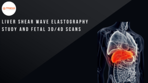
Now, imagine if you could get the same critical information safely, quickly, and without any pain. This is why advanced ultrasound technologies play a vital role in both diagnosis and monitoring. In this blog, we’ll explore the process of Liver Shear Wave Elastography Studies and Fetal 3D/4D Scans. We’ll work on understanding how these advanced imaging methods help in early intervention, improve diagnostic accuracy, and contribute to a healthier liver.
An Overview of Liver Shear Wave Elastography
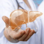
The liver is one of the most essential and metabolically active organs in the human body. This is one of the largest internal organs in the body and plays a central role in maintaining overall health and physiological balance. What makes liver disease particularly challenging is the liver’s ability to withstand significant injury before clinical symptoms become evident.
Liver Shear Wave Elastography, commonly abbreviated as SWE, is an advanced form of ultrasound imaging used to assess the health of the liver in a safe and non-invasive way. Instead of relying on surgery or tissue sampling, this scan measures how stiff the liver tissue is, which is an important indicator of underlying liver damage.
Compared to other liver scans, such as transient elastography, which uses an external vibration to measure liver stiffness, Shear Wave Elastography is built directly into standard ultrasound systems. This allows the scan to be performed in real time alongside routine ultrasound imaging. With this, doctors can carefully select the exact area of the liver to be examined, acquiring more precise results.
From a clinical perspective, SWE is a reliable way to assess liver fibrosis, a condition where healthy liver tissue is gradually replaced by scar tissue due to long-term liver damage. Research has shown a strong relationship between liver stiffness values measured by SWE and the actual degree of fibrosis seen on tissue examination. Detecting liver fibrosis early is critical, as if it is left undetected, it can eventually progress to liver cirrhosis, a serious liver disease that comes with long-term complications.
When is Liver Shear Wave Elastography Recommended?
In everyday medical practice, Shear Wave Elastography is primarily used when doctors need a clear and reliable method to assess the stiffness of the liver. The stiffness of the liver is important for diagnosing liver conditions and planning treatment. Our specialists recommend conducting SWE in the following scenarios:
Chronic Liver Disease Screening: Chronic liver disease (CLD), including viral hepatitis, NAFLD, and alcohol-related liver disease, is common and often develops silently. Liver biopsy has traditionally been used to assess fibrosis, but it is invasive and carries risks such as pain, bleeding, and sampling errors. Shear Wave Elastography (SWE) offers a reliable, non-invasive way to measure liver stiffness and monitor disease.
Fatty Liver Assessment (NAFLD): NAFLD is a leading cause of chronic liver injury. Fibrosis progression strongly impacts long-term outcomes, making early detection crucial. SWE allows clinicians to assess liver stiffness safely, often performing better than conventional ultrasound in identifying fibrosis.
Hepatitis B and C Monitoring: Chronic hepatitis B or C can gradually lead to cirrhosis or liver cancer. Regular assessment with SWE helps track disease progression and response to antiviral therapy, reducing the need for repeated biopsies while providing a safe and painless monitoring option.
Alcohol-Related Liver Issues: Long-term alcohol use can cause progressive liver fibrosis. SWE helps evaluate disease severity, monitor treatment or lifestyle changes, and assess liver health before major procedures, all without the risks of biopsy.
Pre- and Post-Treatment Evaluation: SWE can track changes in liver stiffness before and after interventions. Decreases suggest fibrosis regression, while increases indicate progression, giving clinicians and patients a clear, ongoing picture of liver health.
Who Should Consider Liver Elastography
Our specialists recommend Shear Wave Elastography for the following individuals:
- Individuals with abnormal liver function tests or elevated fibrosis risk.
- Patients with known chronic liver diseases require fibrosis monitoring.
- Individuals who are unable or unwilling to undergo repeated liver biopsies due to risk or discomfort are also included.
- For individuals with obesity or ascites, traditional elastography techniques may fail.
Benefits of Liver Shear Wave Elastography
Compared to other scanning techniques, Shear Wave Elastography offers the following advantages:
Non-Invasive and Repeatable: Unlike liver biopsy, SWE can be repeated as frequently as needed without risk of bleeding or infection.
Simultaneous Anatomical Imaging: Because SWE is integrated with conventional ultrasound, clinicians can choose optimal regions of interest and avoid artifacts, enhancing reliability.
High Diagnostic Accuracy: SWE discriminates between fibrosis stages with a high degree of accuracy, rivaling biopsy for staging clinically relevant fibrosis.
Broad Clinical Utility: Beyond chronic liver disease, SWE also has potential applications in breast, thyroid, and musculoskeletal imaging, reflecting its versatility in evaluating tissue stiffness.
Just as Shear Wave Elastography provides a safe, non-invasive way to assess liver health, modern ultrasound technology also allows expectant parents to observe their baby in remarkable detail without any risk. 3D and 4D fetal ultrasounds extend this principle, offering detailed anatomical and real-time imaging while keeping the process completely non-invasive.
Fetal 3D Ultrasound – What It Shows
Three-dimensional (3D) fetal ultrasound is a safe, non-invasive imaging technique that provides detailed views of a developing baby. By combining multiple two-dimensional scans, 3D ultrasound creates a lifelike image, which adds depth and clarity that standard 2D scans cannot provide.
This technology offers a detailed visualization of fetal anatomy, including facial features, skeletal structures, and overall body contours. In clinical practice, 3D ultrasound can detect congenital conditions, such as cleft lip or subtle structural abnormalities. This is important for clinicians to make early assessments. Above all, it provides expectant parents with a clearer view of their child!
Fetal 4D Ultrasound – Real-Time Imaging
Four-dimensional (4D) ultrasound takes 3D imaging a step further by adding the element of time. This allows clinicians to create a live video of the fetus in the womb. This video is helpful in the observation of movements, expressions, and even early motor activity in real time.
While 2D ultrasound remains the standard for routine prenatal checks, 3D and 4D imaging can offer additional insight when needed. Moreover, these advanced 3D and 4D scans provide a more comprehensive view of fetal development, giving parents a unique glimpse of their baby before birth.
Advantages of 3D/4D Scans for Parents and Doctors
Modern 3D and 4D fetal ultrasounds offer a safe, non-invasive way for clinicians and parents to understand fetal development better. These advanced scans complement traditional 2D imaging by adding depth, clarity, and real-time visualization. Here’s how 3D and 4D scans benefit both medical assessment and parental experience:
Enhanced Anatomical Detail: 3D imaging creates dimensional views of the fetus. This provides doctors with greater spatial resolution for complex growing structures. Such scans can help detect subtle anomalies that may be harder to identify on standard 2D scans.
Real-Time Behavioral Observation: 4D scans add the element of time, producing live videos of fetal movements. Observing behaviors such as limb movements, facial expressions, and early motor activity can help clinicians with insight into neuromuscular development.
Counseling and Parental Bonding: Detailed 3D renderings can be a valuable tool during prenatal counseling, especially when abnormalities are suspected. At the same time, seeing lifelike images or videos of their baby helps parents emotionally connect with the pregnancy, enhancing bonding even before birth.
Together, 3D and 4D ultrasounds combine diagnostic utility with emotional reassurance, making them a powerful addition to modern prenatal care.
Safety of Live SWE and Fetal 3D/4D Imaging Techniques
Both Shear Wave Elastography (SWE) and fetal 3D/4D ultrasounds are generally considered safe when performed by trained medical professionals following established clinical protocols. These non-invasive imaging techniques use non-ionizing ultrasound energy, which does not carry the risks associated with X-rays or other ionizing radiation, making them suitable for repeated assessments when needed.
Liver Shear Wave Elastography: SWE can be used to investigate liver stiffness without requiring tissue sampling or surgery. As the imaging technique relies on safe ultrasound waves, there are no known long-term risks when the procedure is performed correctly. This allows clinicians to monitor chronic liver conditions regularly, track treatment progress, and detect early signs of fibrosis without exposing patients to pain, bleeding, or radiation.
Fetal 3D/4D Ultrasound: Advanced fetal ultrasounds, like 3D and 4D imaging, are clinically recognized as safe for both the mother and the baby. Professional guidelines emphasize the ALARA (As Low As Reasonably Achievable) principle, meaning ultrasound exposure should be minimized in duration and intensity. While these scans are safe when clinically indicated, prolonged scans outside a medical setting are discouraged due to potential risks, such as tissue heating and cavitation.
Overall, when performed by qualified clinicians, both Shear Wave Elastography and fetal 3D/4D ultrasounds provide accurate, non-invasive insights while maintaining a high safety profile.
Preparing for Scans
One of the most significant advantages of non-invasive imaging techniques like Liver Shear Wave Elastography and fetal 3D/4D ultrasound is that preparation is fundamentally simple and stress-free. These scans are designed for patient comfort and do not involve needles, injections, or surgical procedures.
For Liver Shear Wave Elastography: For Liver Shear Wave Elastography, patients are usually advised to fast for a few hours before the scan, as food intake may temporarily affect liver stiffness measurements. Wearing comfortable, loose-fitting clothing can help make the procedure smoother. Your doctor may also review recent blood test results or medical history to interpret the findings accurately.
For fetal 3D/4D Ultrasound: For fetal 3D/4D ultrasound, no special preparation is typically required. However, staying well hydrated in the days leading up to the scan may help improve image clarity. Expectant mothers should carry previous scan reports and follow any specific instructions provided by their clinician.
In both cases, the procedures are quick, painless, and completed within a short time. Patients can resume normal activities instantly after the scan. By following these simple preparation steps, you can ensure accurate results while enjoying a comfortable, anxiety-free scanning experience.
Why Magnus Diagnostics Is the Right Choice for Non-Invasive Imaging
At Magnus Diagnostics, we understand that many patients are anxious about invasive procedures. That’s why we focus on providing non-invasive, pain-free imaging solutions that deliver good results without any discomfort.
Using advanced tools such as Liver Shear Wave Elastography and fetal 3D/4D ultrasound, our team of clinicians can assess liver stiffness, monitor long-term liver issues, and provide detailed images of unborn babies, all in a safe and comfortable environment. These advanced tools in the market allow for early detection, precise evaluation, and better-informed clinical decisions, without needles or surgical procedures!
Magnus Diagnostics combines cutting-edge ultrasound systems with an experienced workforce in the industry to provide a diagnostic experience that is both comfortable and accurate. We offer a wide variety of services like SWE, fetal 3D/4D scans, standard ultrasound, CT, MRI, and lab tests, making it easy and dependable for patients to get all.
Choose Magnus Diagnostics for precision, safety, and compassionate care. Let us help you find accurate results through advanced non-invasive procedures!