Different Types of Scans and Medical Images
Long before the invention and development of X-rays, MRIs, and ultrasounds, physicians and medical personnel relied on the most basic tools available to them – their eyes, ears, and hands! Studies show how ancient healers throughout history would examine the pulse, eyes, and even a patient’s tongue to obtain deeper insights about intestinal ailments. These can be observed as the initial different types of scans conducted by the doctors to help them understand the diseases and medical conditions that occur in the human body in a non-invasive manner.
The medical field had a significant spurt in the 19th century, and the advancements in this era have led to the development of scans and hybrid imaging techniques that pave the way for noninvasive diagnostic imaging by screening not only structural information but also functionality and cellular activity.
Understanding the evolution of scans and medical imaging is crucial, as it reflects how far we’ve come, from reading a pulse with ears and eyes to creating real-time images of organs, blood vessels, and tissues using advanced scanning techniques. In this blog, we will go through the different types of scans and medical imaging processes employed and practiced today and understand their specific purposes and benefits.
Different Types of Scans and Medical Images
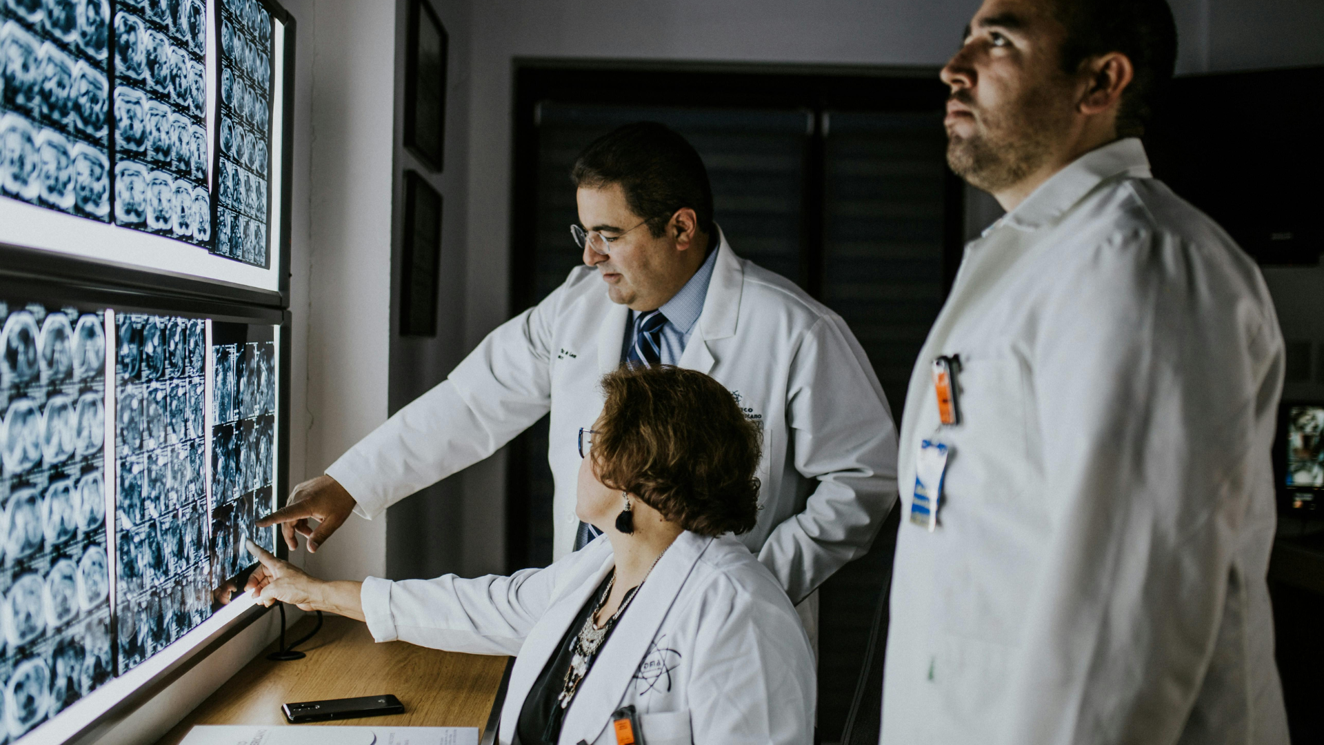
Medical scans and imaging technologies in today’s world have become inevitable tools that empower doctors to non-invasively peek into the human body and acquire details with remarkable clarity. From detecting fractures and tumors to monitoring critical organ functions, medical scans and images also play an instrumental role in guiding doctors through complex surgeries. Medical scans can:
- Detect internal bleeding, fractures, and other injuries.
- Diagnose undetectable illnesses such as cancer, cardiovascular conditions, and other internal infections.
- Monitor ongoing treatments, like results of complex surgeries or chemotherapies, with increased accuracy.
- Assist doctors in complex medical procedures by providing precise data.
While doctors use different types of scans for human body, each has its own definite purpose and offers a unique window into the body to capture the details. Let us take a look at the most commonly used medical scans today and understand how they work and the situations where they are most effective:
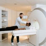
MRI – Magnetic Resonance Imaging
MRI is one of the most advanced and common imaging tools in medicine. This medical scan works by using strong magnetic fields and radio waves to produce highly accurate and detailed 3D images of internal organs and tissues. Unlike other types of scans, such as X-rays or CT scans, MRI does not employ ionizing radiation, making it a safer alternative, especially when repeated scans are required. When it comes to use cases, MRI scanning is employed to detect and monitor neurological conditions such as brain tumors, stroke, multiple sclerosis, nerve injuries, and similar internal issues. This type of scan is the preferred choice by medical professionals when it comes to imaging the spinal cord, joints, muscles, and ligaments for precise results on sports injuries and musculoskeletal disorders. In the department of cardiology, MRIs are used to assess the structural and functional aspects of the human heart. It is also employed in the cases of pregnant women, where radiation is highly dangerous. Doctors also use fMRI (functional magnetic resonance imaging) scans to observe real-time activities inside the body.
Benefits of MRI:
- Highly detailed and high-contrast images can be acquired using contrast agents to acquire details of specific tissues and blood vessels.
- Provides light on details that remain invisible under X-ray and CT scans
- Easier to detect subtle anomalies in the brain, spine, and muscles.
- Can scan any part of the body without the need for employing radiation.
- Non-invasive and relatively safe.
Risks of MRI:
- As MRIs use magnetic fields to acquire the scan, they carry risks for patients with medical implants such as pacemakers, cochlear implants, or older aneurysm clips. The magnetic field may interact and interfere with these devices, leading them to malfunction.
- Individuals with tattoos and permanent makeup may experience burns and skin irritation, as they contain metallic ink.
- MRI scans can be highly uncomfortable for individuals with claustrophobia and may even require sedation.
- The contrast agents used in the scan have been rarely shown to cause allergic reactions.
X-Ray
One of the oldest and most widely used imaging tools, X-rays use low doses of ionizing radiation to acquire 2D images of internal structures inside the body. The scans appear white on the film, as bones absorb X-rays better than the soft tissues, making this type of medical scan easier to detect bone fractures, dislocations, and dental problems. X-rays play an instrumental role when it comes to chest imaging, as they can reveal pneumonia, tuberculosis, fluid buildup, and tumors in the lungs with higher accuracy. The gold standard test to detect breast cancer, mammography, is a specialized form of X-ray. X-rays are also used by medical personnel to discover foreign elements present in the body.
Benefits of X-Ray :
- X-rays are quick, simple, and extensively accessible due to their availability at almost every healthcare center.
- The best option for detecting fractures, chest conditions, and bone density issues.
- Non-invasive and painless procedure.
- Inexpensive compared to other types of scan for human body.
- Useful in emergency situations for rapid and quick diagnosis.
Risks of X-Ray:
- X-rays use ionizing radiation that can increase the risk of cancer in the long run if scans are conducted repeatedly.
- Not useful for acquiring detailed scans of quick tissues.
- The radiation can be highly harmful for developing fetuses, limiting its use in pregnancy.
- The contrast dye used in the process has been shown to cause allergic reactions.
CT/CAT Scan – Computed Tomography
CT scans, commonly referred to as CAT scans, work by combining multiple X-ray images acquired from different angles to produce more detailed cross-sectional views of the body. These scans provide more detailed information compared to standard X-ray scans and are used by doctors to examine bones, blood vessels, and soft tissues in much better detail. CT Scans are widely used to detect internal injuries from trauma, monitor cancers and assess cardiovascular issues with precision. They also assist in biopsies and surgeries. CAT scans are relatively fast and precise medical scans, making them an instrumental diagnostic tool in modern medicine, especially when it comes to emergency rooms and ICUs, where proper and swift diagnosis is essential.
Benefits of CT/CAT Scans:
- CT/CAT medical scans are very effective in acquiring highly detailed cross-sectional images of internal organs and structures in the human body.
- Medical personnel prefer CT/CAT scans for diagnosing cancers, strokes, and trauma-related injuries.
- This type of scan is relatively faster than MRI, making it the go-to option for precise results in case of emergencies.
- Extremely helpful in surgical planning and treatment monitoring.
Risks of CT/CAT Scans:
- Increased exposure to radiation when compared to regular X-rays.
- Not recommended for repeated scans, especially when it comes to children.
- CT/CAT scans usually require contrast dye, which is often found to cause allergic reactions.
- This medical scan cannot acquire as precise soft-tissue images as MRI scans produce.
- Repeated scans that involve high radiation exposure can lead to cancer in the long run.
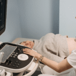
Ultrasound
Ultrasound medical scans use high-frequency sound waves to create images of internal organs and tissues. Ultrasound scans do not use any type of radiation at all, making them extremely safe compared to types of scans such as X-rays and CT/CAT scans. Even though ultrasound scans are mostly associated with pregnancy to monitor the health and growth of fetuses, they are used in the examination of the heart, blood vessels, abdominal organs, and musculoskeletal system. The Doppler ultrasound is an advanced medical scan that can even evaluate blood flow through arteries and veins with high precision, making it a core tool in the department of cardiology and vascular medicine. As it offers real-time data and is non-invasive and safe, ultrasound is one of the most preferred types of scans for human body by medical personnel across the world.
Benefits of Ultrasound:
- Extremely safe, as it uses sound waves and not any type of radiation.
- Ultrasound offers real-time imaging, making it useful in surgeries and biopsies.
- It is easily portable, making it widely available even in smaller clinics.
- Doppler ultrasound makes it easier to obtain detailed evaluations of blood flow and vascular issues.
Risks of Ultrasound:
- Ultrasound is limited, as it cannot provide as detailed images compared to MRI or CT scans.
- The accuracy of the imaging depends on the operator/technician’s skill, contrary to other types of medical scans.
- The results may vary, as sound waves may often not penetrate air-filled spaces and bones. This limitation can often prevent the scan from detecting subtle anomalies and other minute details.
- Ultrasound is not used for in-depth and highly detailed images of organs.
Echocardiogram
An echocardiogram is a specialized ultrasound that has been designed and built to examine the structure and function of the cardiovascular system. These medical scans provide cardiologists with detailed scans of heart chambers, valves, pumping strengths, and blood flows, making them an important tool for identifying diseases related to the heart. Medical personnel around the world have chosen echocardiograms as the standard tool for noninvasive cardiovascular checkups due to their safety, painlessness, and accessibility.
Benefits of Echocardiograms :
- Provides real-time information on cardiovascular functions.
- These scans aid in the detection of valve disorders, heart failure, and congenital heart diseases.
- Assists in monitoring the effectiveness of cardiovascular treatments over time.
- Extremely accessible and cost-effective compared to other types of medical scans.
Risks of Echocardiograms:
- Cannot provide detailed images like MRI and CT scans.
- The quality of scans depends entirely on the technician/operator.
- It is limited when it comes to scanning for obesity or lung diseases.
- Transesophageal echocardiograms can cause throat discomfort due to the invasive nature of the procedure.
Electrocardiograms (ECGs or EKGs)
Commonly referred to as ECGs, electrocardiograms can be defined as tests that record the electrical activity of the heart by using sensors placed across the skin. While ECGs are not imaging scans in the conventional sense, they play a major role in cardiology. ECGs help in detecting small variations in the heartbeats, heart attacks, conduction problems, and electrolyte imbalances in the body. ECGs are the first test conducted by doctors when patients who have chest pain are admitted to the hospital. ECGs are simple, quick, painless, and accurate, making them one of the most critical components in the department of cardiology as well as overall modern medicine.
Benefits of ECG:
- The procedure is quick, painless, and non-invasive.
- Provides immediate results, even in case of emergencies.
- Highly inexpensive and available in most clinics and hospitals.
Risks of ECG:
- Provides data but not in the image format, as acquired through other medical scans.
- It can produce false positives and negatives.
- This medical scan may miss certain heart diseases.
- The interpretation of data depends on the knowledge and skills of the medical personnel.
PET Scan – Positron Emission Tomography
PET scans are advanced imaging tests that provide insights using data acquired from cellular and metabolic activities inside the body. This medical scan works by revealing the functions of tissues and organs, unlike other types of scans, which focus on the structures. These scans are highly effective for cancer detection due to the fact that cancer cells show higher activity. PET scans work by injecting a radioactive substance called a radiotracer that accumulates in areas where cancer cells or cancerous growth are present. PET scans are also used in the diagnosis of brain disorders such as Alzheimer’s, epilepsy, and Parkinson’s disease. PET scans are often combined with other scans to acquire both structural and functional data, making them one of the most important medical scans on the market.
Benefits of PET Scans:
- PET scans help in detecting metabolic changes before they start affecting anatomical functions.
- They are highly effective in identifying cancerous tumors as well as in monitoring the treatment responses.
- This medical scan can also be used to assess cardiovascular functions and proper blood flow.
- Combining PET with CT/CAT and MRIs provides extensive accuracy.
Risks of PET Scans:
- Individuals are exposed to radiation through the radiotracers.
- These scans are limited in availability and also cost a lot.
- The presence of radioactive substances makes it ill-suited for pregnant and breastfeeding women.
- The radiotracer injections can cause allergic reactions.
Why Medical Scans are Important for Doctors
Today, medical scans stand as the vital tools that assist healthcare providers in diagnosing and monitoring a diverse range of medical conditions. These different types of scans offer detailed images that help in identifying abnormalities, planning treatments, and monitoring the progress of the treatment in a non-invasive manner.
Understanding the different types of medical scans, their uses, benefits, and drawbacks is important for patients to make informed decisions when it comes to their healthcare. While each type of scan has its own specific applications, they wholly contribute to the medical field in diagnosing and treating medical conditions effectively.