Functional MRI (fMRI): A Modern Window into the Brain
The brain is a complex organ, and with modern imaging technology, we can look inside it now like never before. One such groundbreaking technique is functional MRI, commonly known as fMRI. It is used in clinical diagnostics and psychological research to revolutionise observing and comprehending brain activity.
Throughout this article, we will cover the meaning of fMRI, what it is, how it functions, its advantages and disadvantages, and why it has become so important in both medicine and neuroscience.
What is Functional MRI?
The fMRI full form is functional Magnetic Resonance Imaging. It is a non-invasive imaging technique that measures and maps out the brain function. Unlike traditional MRI, which captures static images of the brain’s anatomy, functional MRI tracks changes in blood flow related to neural activity in real time.
This is based on the fact that the more active a particular portion of the brain is, the more oxygen is needed. The scanner records these variations in blood oxygenation and flow, which can be visualised by clinicians and researchers to help understand areas of the brain that are being utilised when carrying out different mental or physical activities.
How Common are fMRI scans?
While fMRI scans are not as commonly used as standard MRI scans, they are increasingly utilised in both clinical and research settings. In hospitals and sophisticated medical diagnostics facilities such as Magnus Diagnostics, fMRI is generally prescribed in cases of:
- Pre-operative surgery planning in patients with brain tumours and epilepsy.
- Studying the brains of people with neurological disorders.
- Cognitive and behavioural Research.
- Investigating mental disorders such as depression, schizophrenia, or autism.
With advancements in functional MRI (fMRI) radiology, the demand for fMRI is steadily increasing, particularly as the medical community places greater emphasis on understanding brain function in conjunction with its structure.
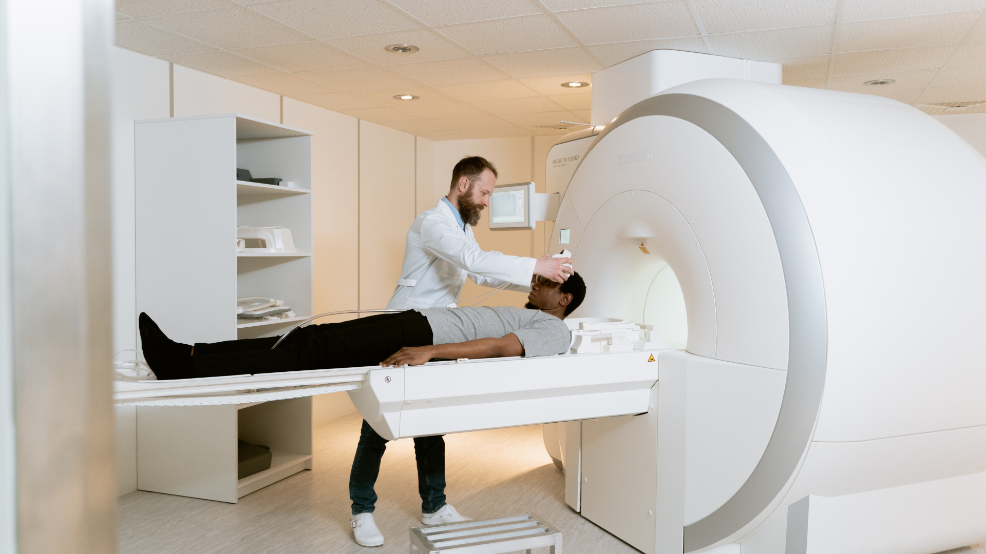
How is the Procedure Performed?
An fMRI procedure is very similar to a regular MRI except for a few minor adjustments. A high-field MRI machine is used to scan, and it is mostly a 1.5 Tesla or 3 Tesla in strength.
Generally, here is a stepwise overview of what is expected:
Preparation:
You may be asked to put on a hospital gown and remove any metal items, such as jewellery or glasses.
Positioning:
You will then lie flat on a motorised table, which will slide into the cylindrical tunnel of the MRI scanner. Small cushions may be placed around your head to keep it still.
Task Performance (possibly):
Some types of functional MRI involve activation of brain regions. You will be asked to do something, such as tapping fingers, solving puzzles, reading, or looking at pictures.
Imaging Acquisition:
The scanner, together with multiple acquisitions, detects changes in the blood oxygen levels. On the whole, it is about 30 minutes to 60 minutes long, depending on the length of the scan.
You will be continuously monitored by either a radiologist or an MRI technologist in the control room adjacent to the scanner.
What Will I Experience During and After the fMRI?
You will hear loud knocking or tapping sounds during the scan as the machine captures images. Usually, earplugs or headphones will be provided to keep you comfortable. You must remain as motionless as possible to obtain clear images.
The scan is painless, and generally, no contrast dye is needed. After the scan, you can resume your normal activities. No recovery time or special care will be necessary whatsoever.
Advantages and Disadvantages of fMRI
As with all medical technology, this functional MRI comes with its pros and cons.
Pros
- Non-invasive and safe: No radiation involved.
- Spatial resolution: Excellent pinpointing of active brain regions.
- Real-time visualisation of brain activity: Helps surgeons and researchers.
- Versatility: Applications stretch across neurology, psychology, psychiatry, and cognitive science.
Cons
- Expensive: fMRI is costlier than other imaging techniques such as EEG.
- Highly sensitive to movement: Even a slight movement distorts images.
- Limited availability: Only a handful of imaging centres provide fMRI scans.
- Indirect measure: It measures blood flow change and not electrical activity directly.
Conclusion
In the modern era, fMRI stands at the forefront of diagnostic and research tools in brain science. From presurgical planning and performing neurology examinations to psychological studies, fMRI provides a singular non-invasive way of observing the brain in action.
As technology advances further, and imaging of this calibre continues becoming accessible in centres like Magnus Diagnostics, it is all the more promising to witness improved diagnosis, treatment planning, and understanding of the human mind.
If you were advised to undergo an fMRI scan, rest assured that you have one of the most advanced applications available in medicine today.
If you’re looking for a reliable MRI scan in Thrissur, Magnus Diagnostics is the place to go. They offer accurate and high-quality scans to help with both medical and research purposes.
Frequently Asked Questions
1. What is the role of MRI in the brain?
MRI, especially fMRI, is also necessary for diagnosing and understanding brain-related disorders. While standard MRI helps visualise structural problems like tumours or lesions, fMRI shows what parts of the brain are active when someone is performing a task or resting. It is therefore useful in pre-surgical planning, monitoring recovery processes after stroke or injury, in questioning unexplained symptoms, and in adding to the enquiry of a psychiatric nature.
2. Why is fMRI better than EEG?
fMRI and EEG both study brain activity, but in different ways. EEG records electrical changes and detects rapid changes in brain function, but lacks spatial resolution. The fMRI, on the other hand, measures changes in blood flow and therefore possesses a higher spatial resolution that can show the exact location of brain activity. EEG is great at capturing very fast responses, whereas fMRI excels at determining what parts of the brain are active during different functions such as language or movement. Both are non-invasive; however, when it is critical to know exactly where the activity occurs, fMRI takes precedence.
3. What are the strengths and limitations of fMRI?
Strengths
- Detailed anatomical images.
- Enables the understanding of functional brain networks.
- Allows for the early diagnosis of neurological and psychiatric conditions.
- Supports neuroscience and cognitive research.
Limitations
- Expensive and time-consuming.
- It cannot be used on patients with metal implants, such as pacemakers.
- Not recommended for severely claustrophobic individuals.
- Sometimes fails to capture very rapid neural responses.
The result can vary depending on the task or the scanner used.
4. Why use fMRI in psychology?
Functional MRI has become the staple of research in psychological and cognitive fields. It motivates scientists to investigate the neural correlates of mental processes, such as:
- Decision making.
- Memory recall.
- Emotional regulation.
- Language comprehension.
- Attention and perception.
By giving information regarding which brain areas are activated during an activity involving mental processes, fMRI helps understand how cognition, feelings, and behaviour are represented in the brain. It is also useful in examining how brain function varies in people with mental disorders such as depression, ADHD, PTSD, and schizophrenia.