Pediatric MRI: Procedure, Preparation, and Safety Explained
When it comes to diagnosing health conditions in children, accuracy and safety are always top priorities. Modern medical imaging techniques play a key role in the timely identification of health issues, guiding treatment, and ensuring proper follow-up. Among the various imaging tools for children, Magnetic Resonance Imaging (MRI) is noted for its detailed and radiation-free imaging capabilities.
When an MRI is performed on children or infants, it’s known as a pediatric MRI or baby MRI scan. It is a specialised procedure that uses powerful magnetic fields, radio waves, and computer technology to create detailed images of the organs, tissues, and structures inside a child’s body.
In Thrissur, where access to advanced healthcare and diagnostic facilities continues to grow, pediatric MRI plays an important role in helping doctors diagnose and manage conditions accurately and safely. In this blog, we will cover everything you need to know about pediatric MRI, including its procedures, preparation, and safety considerations.
What is Pediatric MRI?
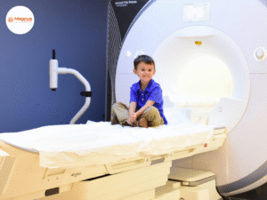
A pediatric MRI is a type of MRI scan specifically designed for infants and children. It uses a strong magnetic field, radio waves, and advanced computer technology to produce highly detailed images of the inside of the body. Unlike X-rays or CT scans, MRI does not use radiation, making it especially suitable for children.
Doctors may recommend a pediatric MRI to examine the brain, spine, heart, abdomen, joints, or other organs. The scan helps in diagnosing conditions such as developmental delays, congenital abnormalities, infections, injuries, or tumours.
What is the Procedure of a Pediatric MRI Scan?
A pediatric MRI scan is a simple and painless procedure, but it involves careful preparation and patience. To ensure detailed imaging while keeping the child comfortable, a pediatric MRI scan typically involves several steps:
- Preparation: Preparing the child for the procedure is the first step. It involves explaining the process in a friendly, age-appropriate way so that the child feels less anxious. All metallic objects, such as jewellery, clips, or watches, are removed because MRI uses a powerful magnetic field. If a contrast-enhanced MRI is required, a small intravenous (IV) line may be inserted to administer a special dye that helps highlight certain tissues more clearly.
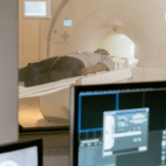
- Positioning the child: The child is then asked to lie down on a narrow, movable table. Straps, bolsters, or cushions are used to keep the child still and prevent any movement during the scan, as even small motions can blur the images. The table slowly moves into the scanner’s tunnel to start the procedure. Specialised coils are placed around or near the body part being imaged. These coils help capture high-quality pictures by transmitting and receiving radio signals.
- Scanning: Once the procedure begins, the MRI machine makes tapping or knocking sounds as it takes images. This is completely normal. To protect the child from the noise, earplugs or headphones are provided. The technologist monitors the child closely from an adjacent control room and communicates through an intercom. Depending on the safety policy of the diagnostic centre, parents may be allowed to stay inside the room for reassurance.
- Post-Scan Care: Once the imaging is complete, the table moves out of the scanner, and the technologist helps your child sit up or get off the table carefully. If sedation was used, your child will be moved to a recovery area, where medical staff will observe them until they are fully awake and alert. Minor drowsiness or disorientation after sedation is normal and wears off within a short time.
- Image Review and Reporting: The captured images are then reviewed by a pediatric radiologist, who interprets them and prepares a detailed report. This report is shared with your child’s doctor, who will explain the findings and discuss any further steps or treatments needed.
How to Prepare the Child for the MRI Scan?
Unlike adult MRI scans, a pediatric MRI is often more challenging, not because of the procedure itself, but due to the need for the child’s cooperation. Since the experience can feel unfamiliar and even intimidating to children, they may become anxious or restless. Therefore, proper preparation and reassurance beforehand can greatly help ensure the procedure goes smoothly and is completed successfully.
Explain the process to the child in a simple and trusting manner to ease their anxiety. If the scan requires sedation or fasting, follow all instructions given by the diagnostic centre carefully. Typically, fasting is required for a few hours before the procedure to ensure safety during sedation.
The child may be asked to wear a hospital gown and remove any metal objects to ensure safety during the scan. It’s best to dress your child in loose, metal-free clothing and remove all accessories, hairpins, and watches beforehand.
Are There Any Alternatives to a Pediatric MRI Scan?
An MRI scan is one of the most advanced and detailed imaging techniques and is primarily chosen for its effectiveness and safety. However, there are a few alternative imaging techniques the doctor might consider for your child.
- Ultrasound: Ultrasound is often the first imaging option for infants because it’s quick, non-invasive, and uses sound waves instead of radiation. It’s particularly useful for examining soft tissues, the abdomen, and even the brain in newborns through the soft spot (fontanelle). However, ultrasound provides less detailed images than MRI and may not be sufficient for deeper or more complex structures.
- CT Scan: A CT scan (Computed Tomography) uses X-rays to create detailed cross-sectional images of the body. CT scans are faster and may be used in emergencies, especially for assessing bones or internal bleeding.
- X-Rays: X-rays are used primarily for bone injuries or chest assessments. While they are useful for quick diagnosis, they lack the comprehensive detail and tissue contrast that MRI offers.
That being said, it is important to note that an MRI scan is often the right option because of its precision and superior image quality.
Are There Any Risks Involved in Pediatric MRI?
MRI is widely regarded as a very safe imaging procedure, especially for children. Since it uses magnetic fields and radio waves instead of radiation, it poses no long-term health risks.
While there are no known risks for pediatric MRI, discomfort and anxiety can be a problem for children undergoing the procedure. The machine’s loud sounds and confined space can be unsettling, especially for young children. This is why most pediatric centres use ear protection, distraction methods, or mild sedation when necessary.
Another possible risk involves the use of contrast dyes. When a contrast dye is used for enhancing specific organs or tissues, there is a very small risk of an allergic reaction. The dye, called gadolinium, is generally safe and well-tolerated. Rarely, children might experience mild reactions such as hives, itching, or wheezing.
If sedation or anaesthesia is required, there may be minor, temporary side effects like grogginess, irritability, or nausea. These effects usually pass within a few hours. During and after sedation, your child will be carefully monitored by trained professionals to ensure full safety.
In short, no major risks or complications are usually experienced during or after the procedure. Since the scan is performed under strict safety protocols by expert radiologists and technologists, you can be assured that your child’s well-being is always the top priority.
What Happens After the Scan?
Once the MRI scan is complete, a technologist or nurse will help your child off the table. In most cases, children can go home right away and resume their normal activities. If sedation was used, your child will be moved to a recovery area, where medical staff will monitor them until they are fully awake and alert.
It’s common for children to feel a little sleepy or irritable after sedation, but this usually passes within a few hours. Once they’re awake, encourage them to drink fluids and have a light meal. Before you leave, the medical team will provide clear post-procedure care instructions to follow at home.
The MRI images will be reviewed by a radiologist, who prepares a detailed report and sends it to your child’s referring doctor, who will then discuss the results with you. They’ll explain what the findings mean and whether any further tests or treatments are needed.
Why Choose Magnus Diagnostics for Precise Diagnostic Services in Thrissur?
Selecting the right diagnostic centre is the primary step in getting an accurate medical diagnosis and effective treatment. With the continually advancing healthcare standards in Thrissur, patients deserve diagnostic services that combine precision, efficiency, and compassion.
At Magnus Diagnostics, we focus on delivering accurate results with utmost care and efficiency. Our aim is to make every diagnostic experience smooth, reliable, and reassuring for every patient.
Here is what sets us apart:
- Comprehensive Diagnostic Services: At Magnus Diagnostics, we offer a wide range of diagnostic services, from lab tests to imaging. Whether you need an MRI, CT scan, or routine blood work, our comprehensive facility ensures convenience and continuity of care.
- Expertise: With a team of experienced radiologists, pathologists, and certified technicians, we prioritise accuracy and patient safety. With years of clinical expertise, they interpret every test with care, ensuring your reports are clear, precise, and dependable.
- Advanced Equipment: We use advanced imaging and laboratory technologies to deliver precise results. With reliable technology, we help medical experts detect conditions earlier, plan better treatments, and make confident medical decisions.
- Quick Turnaround Time: We understand how important timely results are. That’s why we focus on fast reporting without compromising on quality.